Gradient vector analysis-based retinal image micro-aneurysm automatic detection and identification method

A gradient vector and automatic detection technology, which is applied in image enhancement, image analysis, image data processing, etc., to achieve easy operation and improve the effect of detection and recognition

Inactive Publication Date: 2017-05-31
HARBIN INST OF TECH
View PDF0 Cites 11 Cited by
  • Summary
  • Abstract
  • Description
  • Claims
  • Application Information

AI Technical Summary

Problems solved by technology

Therefore, it is still a very challenging task to automatically detect and identify this tiny target from fundus images.

Method used


Image

  • Gradient vector analysis-based retinal image micro-aneurysm automatic detection and identification method
  • Gradient vector analysis-based retinal image micro-aneurysm automatic detection and identification method
  • Gradient vector analysis-based retinal image micro-aneurysm automatic detection and identification method

Examples


specific Embodiment approach 1

[0026] Specific Embodiment 1: This embodiment discloses a method for automatic detection and recognition of retinal image microaneurysms based on gradient vector analysis. The method includes the following steps:

[0027] Step 1: Candidate microaneurysm detection; first microaneurysm extraction, the microaneurysm extraction mainly includes three steps of vessel removal, candidate microaneurysm location and segmentation;

[0028] Step 2: Extraction of candidate microaneurysm features;

[0029] Step 3: Microaneurysm identification.

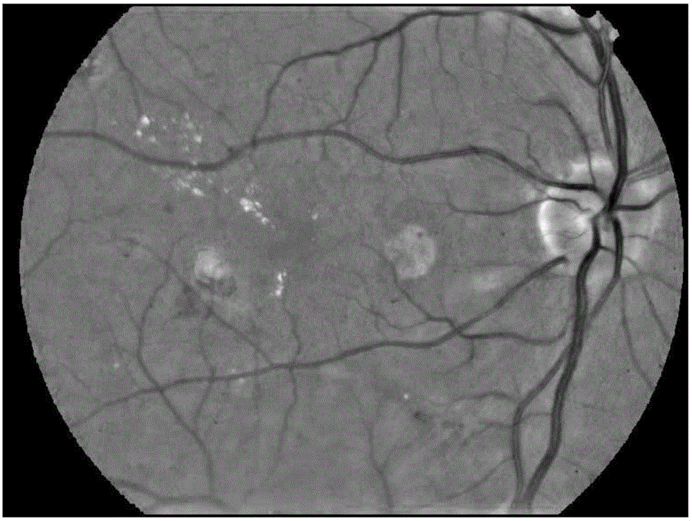
[0030] Considering that the microaneurysm has the greatest contrast with the background in the green channel of the retinal image, the green channel will be used as the input image for subsequent operations. Before extracting candidate microaneurysms, in order to remove the noise of the fundus image while preserving the edge of the arteriole, the image was smoothed by edge-preserving filtering based on the weighted least squares framework. The sha...

specific Embodiment approach 2

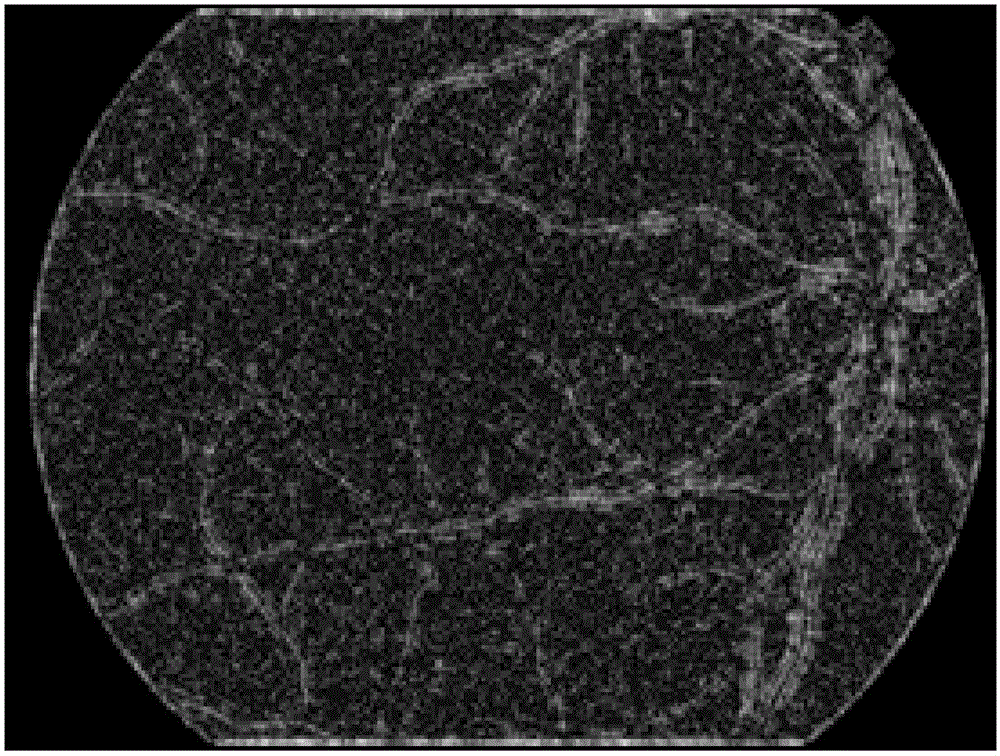
[0031] Specific embodiment two: the method for automatic detection and recognition of retinal image microaneurysms based on gradient vector analysis described in specific embodiment one, in step one, the described blood vessel removal method is: by analyzing the different gradients between microaneurysms and blood vessels Distribution characteristics, to achieve the removal of blood vessels, and retain the real microaneurysms while removing blood vessels; the gradient vector x, y components are regarded as two random variables, by calculating and analyzing the correlation of all gradient vector components in the target area Variance matrix; for retinal vessels, the eigenvalues ​​of the gradient component covariance matrix have a dominant eigenvalue; while for arterioles, the corresponding eigenvalues ​​are approximately equal.

[0032] The capillaries in which microaneurysms are located are not visible in fundus retinal images; therefore, microaneurysms are usually isolated fro...

specific Embodiment approach 3

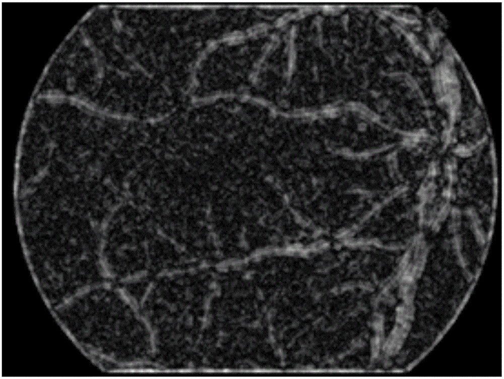
[0040] Specific embodiment three: the method for automatic detection and identification of retinal micro-targets (microaneurysms) based on gradient vector analysis described in specific embodiment one, in step one, the method for the location of candidate microaneurysms is: by calculating The second derivatives of candidate microaneurysms in multiple directions can be used to precisely locate microaneurysms.

[0041] After removing blood vessels, there will still be some tiny blood vessel fragments, background noise, and other fundus dark circular structures. Next, the present invention will locate candidate microaneurysms in the blood vessel removal image, while suppressing the appearance of non-microaneurysms. Considering that the above-mentioned non-microaneurysms and microaneurysms have similar gray values, it is not feasible to directly use gray-scale information to locate candidate microaneurysms.

[0042] Because the microaneurysm has a Gaussian shape distribution in g...


PUM


Abstract

The invention discloses a gradient vector analysis-based retinal image micro-aneurysm automatic detection and identification method. The method comprises the following steps of 1, detecting a candidate micro-aneurysm: firstly extracting the micro-aneurysm mainly by three steps of removing blood vessels, locating the candidate micro-aneurysm and segmenting the candidate micro-aneurysm; 2, extracting features of the candidate micro-aneurysm; and 3, identifying the micro-aneurysm. A new gradient vector analysis-based retinal image micro-aneurysm automatic detection and identification method is proposed in combination with a sample unbalanced classifier through analyzing gradient vector distribution situations of different dark targets in a fundus retinal image. The method also can be effectively applied to the detection of a tiny foreign matter object in an infusion image, the detection of a tiny infrared target in a remote sensing image and the detection of a tiny defective target in a workpiece surface image.

Description

technical field [0001] The invention relates to a method for automatic detection and identification of retinal image microaneurysms. Background technique [0002] Tiny targets in retinal images, such as microaneurysms (MAs), can provide effective auxiliary information for the identification of patients with fundus diseases. They usually appear as dark red dot-shaped targets in retinal images, and some microaneurysms are difficult to distinguish from the background. In addition, some microaneurysms were irregular in shape, clustered together, or near retinal vessels. Therefore, it is still a very challenging task to automatically detect and recognize this tiny target from fundus images. Therefore, the present invention proposes a method for automatic detection and identification of retinal microaneurysms based on gradient vector analysis. The detection of tiny targets in retinal images has important reference value for the detection of tiny foreign objects in infusion imag...

Claims


Application Information

Patent Timeline
Patent Type & Authority Applications(China)
IPC IPC(8): G06T7/00G06T7/10G06K9/54
CPCG06T7/0012G06T2207/30101G06T2207/30096G06T2207/30041G06T2207/20081G06T2207/10004G06V10/20
Inventor 邬向前卜巍戴百生
Owner HARBIN INST OF TECH
Why Eureka
Try Eureka