Medical image display method and device

A medical image and display method technology, applied in the medical field, can solve problems such as prolonging the required time, increasing the workload of staff, and relying on staff for volume data and MPR images

Active Publication Date: 2017-05-31
SHANGHAI UNITED IMAGING HEALTHCARE
View PDF9 Cites 3 Cited by
  • Summary
  • Abstract
  • Description
  • Claims
  • Application Information

AI Technical Summary

Problems solved by technology

[0006] The purpose of the present invention is to provide a medical image display method and device to solve the problem that using the existing medical image display method and device to reconstruct volume data and MPR images of each time phase depends on the staff, resulting in increased workload of the staff , prolonging the time required for TAVI preoperative assessment

Method used


Image

  • Medical image display method and device
  • Medical image display method and device

Examples


Embodiment 1

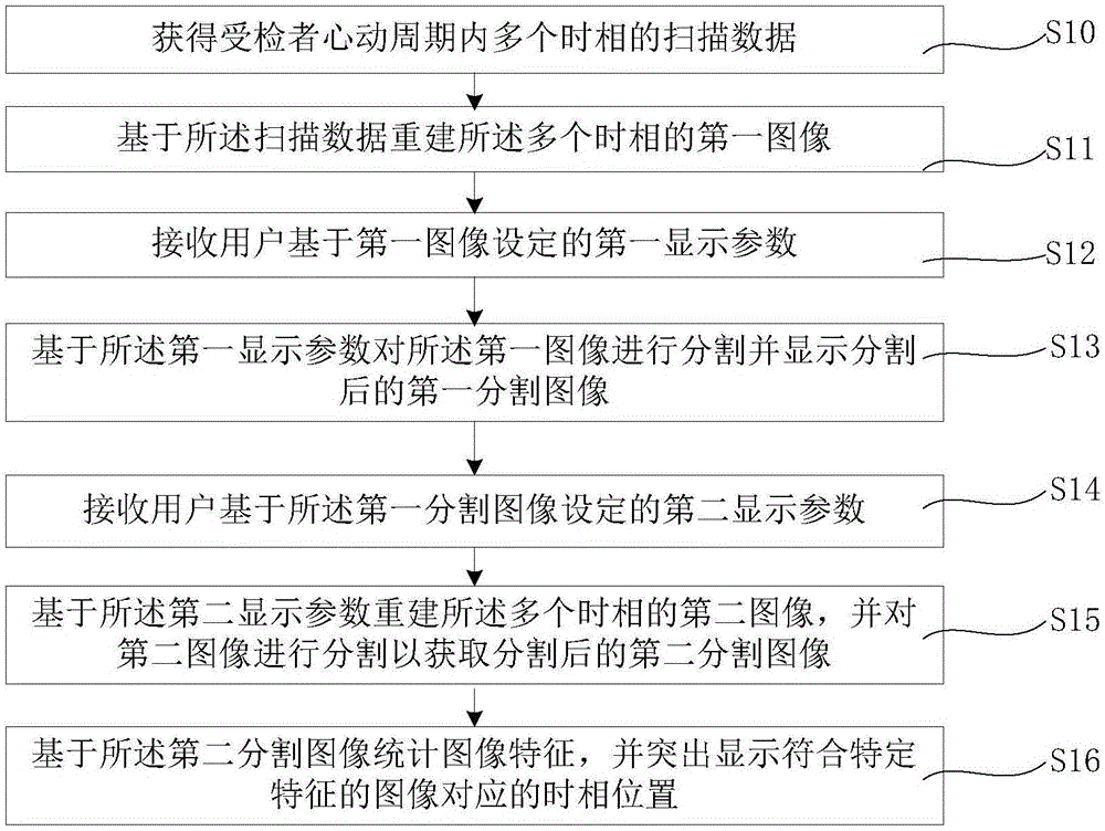
[0034] Please refer to figure 1 , which is a flowchart of the medical image display method in this embodiment, and the medical image display method and device mainly include the following steps:

[0035] First, step S10 is performed to obtain scanning data of multiple phases in the cardiac cycle of the subject;

[0036] Preferably, MDCT is used for scanning to obtain scanning data of multiple time phases within the subject's cardiac cycle. Specifically, the ways of using MDCT to scan and collect data include but are not limited to the following two ways: MDCT uses an electrocardiogram (EGC)-gated retrospective spiral acquisition method to collect data, or MDCT uses an ECG-triggered gated acquisition method to collect data. data collection.

[0037] Next, step S11 is performed to reconstruct the first images of the plurality of time phases based on the scan data;

[0038] Next, step S12 is performed to receive the first display parameters set by the user based on the first i...

Embodiment 2

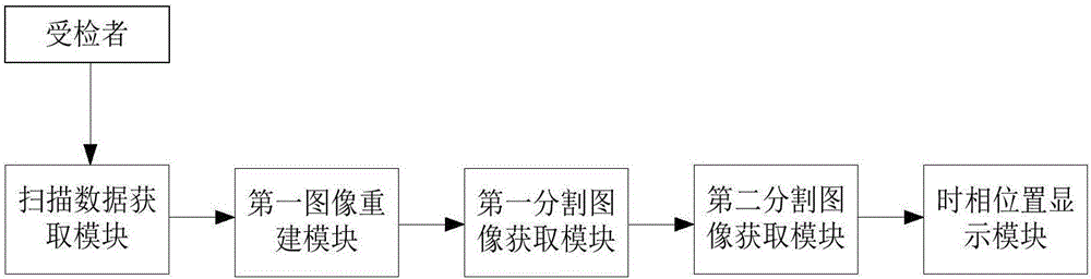
[0062] Please refer to figure 2 , which is a schematic structural diagram of the medical image display device in this embodiment. like figure 2 As shown, the medical image display device includes: a scan data acquisition module, a first image reconstruction module, a first segmented image acquisition module, a second segmented image acquisition module and a phase position display module; a scan data acquisition module for acquiring Scan data of multiple time phases in the cardiac cycle of the examiner; the first image reconstruction module is used to reconstruct the first images of the multiple time phases based on the scan data; the first segmented image acquisition module is used to receive The first display parameter set by the user based on the first image, and based on the first display parameter, the first image is segmented and the segmented first segmented image is displayed; the second segmented image acquisition module is used for receiving The user reconstructs ...


PUM


Abstract

The invention provides a medical image display method and device. The medical image display method comprises the following steps of: obtaining multi-time-phase canning data of an examined person in a cardiac cycle; re-establishing a first multi-time-phase image based on the scanning data; receiving a first display parameter set by a user based on the first image; based on the first display parameter, segmenting the first image, and displaying a first segmented image after segmentation; receiving a second display parameter set by the user based on the first segmented image; re-establishing a second multi-time-phase image based on the second display parameter, and segmenting the second image to obtain a second segmented image after segmentation; and, counting image features based on the second segmented image, and highlighting the time phase position corresponding to the image according with specific features. On the basis of the medical image display method provided by the invention, multi-time-phase data and a three-dimensional MPR image of the examined person can be automatically re-established according to evaluation requirements of doctors; and thus, manual re-establishing of users and the heavy and complicated multi-time-phase image adjusting workload can be avoided.

Description

technical field [0001] The present invention relates to the field of medical technology, and in particular, to a medical image display method and device. Background technique [0002] Transcatheter aortic valve implantation (TAVI) is a new minimally invasive valve replacement surgery developed in recent years. Before performing TAVI, an accurate preoperative assessment of the patient's aortic valve annulus and valve conditions and other cardiovascular structures is required, and then the prosthetic valve replacement and surgical plan are selected according to the results of the assessment. [0003] Multidetector-row CT (MDCT), as an important imaging method for preoperative evaluation of TAVI, mainly uses MDCT imaging with high temporal and spatial resolution, which can clearly display the aorta and other cardiovascular structures. The characteristics of three-dimensional multi-planar reconstruction (MPR) images can be reconstructed. [0004] The evaluation result of the o...

Claims


Application Information

Patent Timeline
IPC IPC(8): G06T17/00G06T7/10G06F19/00
CPCG06F19/321G06T17/00G06T2207/10081G06T2207/30048
Inventor 郝欣
Owner SHANGHAI UNITED IMAGING HEALTHCARE
Why Eureka
Try Eureka