
A kind of blood vessel imaging method

A technology of vascular imaging and final image, applied in medical science, diagnosis, diagnostic recording/measurement, etc., can solve the problems of long echo time, large coding gradient, and affecting image results, etc., and achieve the effect of shortening echo time

Active Publication Date: 2021-03-09
SHANGHAI JIAO TONG UNIV
View PDF6 Cites 0 Cited by
  • Summary
  • Abstract
  • Description
  • Claims
  • Application Information

AI Technical Summary

Problems solved by technology

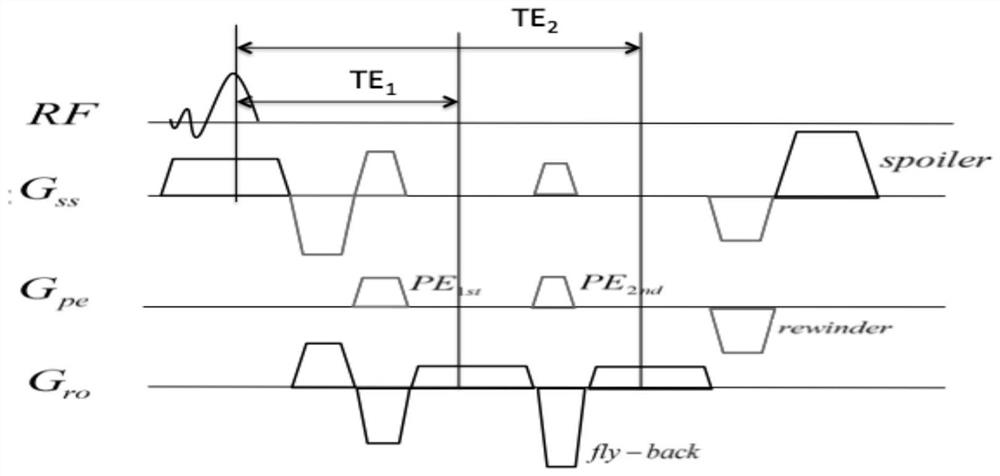
However, there are three main problems in this three-dimensional time-flight vascular imaging double-echo sequence: (1) The echo time (effective TE) of the first echo is relatively long due to the large coding gradient in the layer selection direction; 2) The information at the four corners of the k-space edge is relatively redundant, which increases additional scanning time; (3) Due to the phase modulation of the double echo, artifacts are introduced in the phase direction, which affects the final image result

Method used


Image

  • A kind of blood vessel imaging method
  • A kind of blood vessel imaging method
  • A kind of blood vessel imaging method

Examples


Embodiment Construction

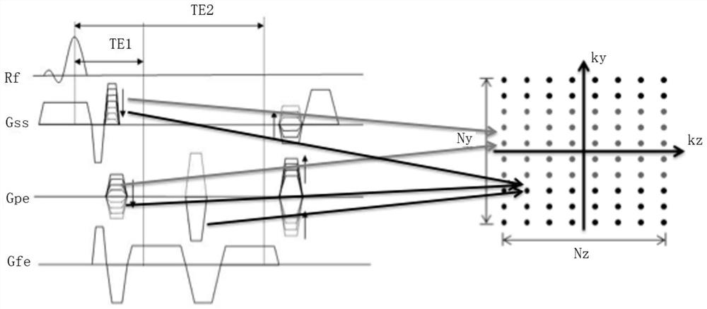
[0042] Such as figure 1As shown in the prior art, the three-dimensional time-of-flight method for vascular imaging double-echo sequence, and the corresponding k-space filling schematic diagram. The gray dot represents the first echo signal (sampling time at TE1), and its position in k-space is determined by the layer-selected phase encoding and phase encoding gradient before the first echo. The black dot represents the second echo signal (sampling time at TE2), and its position in k-space is determined by all phase encodings before the second echo signal and layer-selected phase encodings before the first echo.

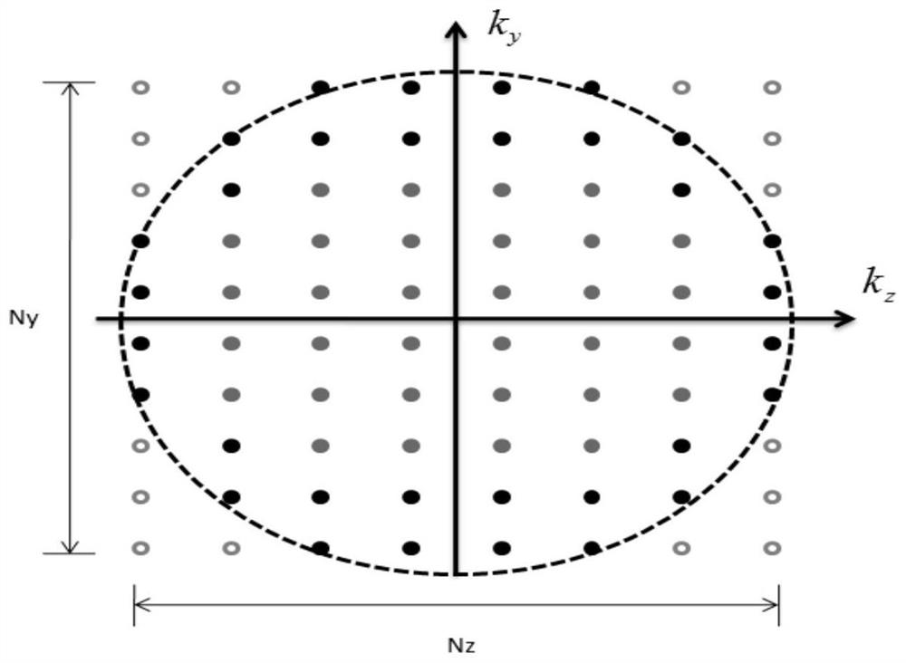
[0043] figure 2 It is a schematic diagram of k-space filling corresponding to the double-echo sequence of vascular imaging by the three-dimensional time-flight method based on the k-space from the outer to the inner ellipse center trajectory. Hollow points at the four corners are not acquired and are zero-filled during reconstruction to obtain the final image. The...


PUM


Abstract

The invention discloses a three-dimensional time-flight double-echo sampling method for vascular imaging, relates to the field of magnetic resonance imaging, and comprises the following steps: y ‑k z Points in the space are divided into points in the ellipse area and points outside the ellipse area; the points in the ellipse area are divided into two parts according to the distance from the center point of k-space, and the first echo and the second echo are filled respectively. The echo signal of the first echo with a shorter echo time is filled in a position close to the center point of k-space, and the echo signal of the second echo with a longer echo time is filled in a position far from the center point of k-space; The k-space positions in the sequence correspond to the different phase directions on the sequence and the gradient encoding of the layer-selected phase direction; the collected data are filled into their corresponding k-space positions, and the positions outside the ellipse area are filled with zeros for reconstruction. The sampling method of the present invention shortens the scanning time and the echo time on the basis of the double-echo sampling scheme, avoids artifacts caused by human body movement, and improves the effect of blood vessel imaging.

Description

technical field [0001] The invention relates to the field of magnetic resonance imaging, in particular to a blood vessel imaging sampling method. Background technique [0002] Magnetic resonance angiography (magnetic resonance angiography, MRA) has become one of the routine techniques for MR blood vessel examination. Compared with digital subtraction angiography (digital subtraction angiography, DSA), it has the advantages of non-invasive, simple, low cost, and no contrast agent. . Different from other vascular imaging methods, MRA technology can not only provide morphological information of blood vessels, but also provide quantitative information such as blood flow direction, flow velocity, and flow rate. [0003] At present, many MRA methods have appeared clinically, and the three-dimensional time-of-flight angiography (3D TOFMRA) sequence is one of the important methods widely used in clinical practice, and its principle is the inflow effect. In order to meet the clinic...

Claims


Application Information

Patent Timeline
Patent Type & Authority Patents(China)
IPC IPC(8): A61B5/055
CPCA61B5/055A61B5/489A61B5/7203A61B5/7207A61B5/7235
Inventor 杜一平陈名亮
Owner SHANGHAI JIAO TONG UNIV
Learn More