A Polypeptide Liposome Capable of Morphological Transformation in Lysosomes of Tumor Cells

A technology of tumor cells and lysosomes, which is applied in the field of polypeptide self-assembly nanosystems, can solve the problems of stimulation sensitivity and achieve good specificity, enhanced cytotoxicity, and high drug release efficiency

Active Publication Date: 2021-06-22
THE NAT CENT FOR NANOSCI & TECH NCNST OF CHINA
View PDF2 Cites 0 Cited by
  • Summary
  • Abstract
  • Description
  • Claims
  • Application Information

AI Technical Summary

Problems solved by technology

In addition, the lack or decrease in activity of certain phospholipases in lysosomes in tumor cells makes lysosomes fragile and sensitive to stimulation

Method used


Image

  • A Polypeptide Liposome Capable of Morphological Transformation in Lysosomes of Tumor Cells
  • A Polypeptide Liposome Capable of Morphological Transformation in Lysosomes of Tumor Cells
  • A Polypeptide Liposome Capable of Morphological Transformation in Lysosomes of Tumor Cells

Examples


Embodiment 1

[0030] L-amino acids protected by benzoic acid and Fmoc (Fmoc-Lys(Boc)-OH, Fmoc-Asp(tBu)-OH, Fmoc-Gly-OH, Fmoc-Ala-OH, purchased from Jill Biochemical (Shanghai) Co., Ltd. ) as raw material, on dichlorotrityl chloride resin (purchased from Jill Biochemical (Shanghai) Co., Ltd.), in the presence of coupling reagents and cleavage reagents commonly used in this field, prepared on the resin by solid phase synthesis Polypeptide (the main body is 6 alanines, the C-terminal of the main body is connected to the tumor cell targeting peptide RGD, and the N-terminal of the main body is modified with a benzene ring). After a simple cleavage reaction, the peptide is separated from the resin, and the pure white peptide powder is obtained after subsequent precipitation, washing, and drying.

[0031] The specific experimental process is as follows:

[0032] First, take 1.01 g of dichlorotrityl chloride resin (purchased from Jill Biochemical (Shanghai) Co., Ltd.) to the peptide synthesis devi...

Embodiment 2

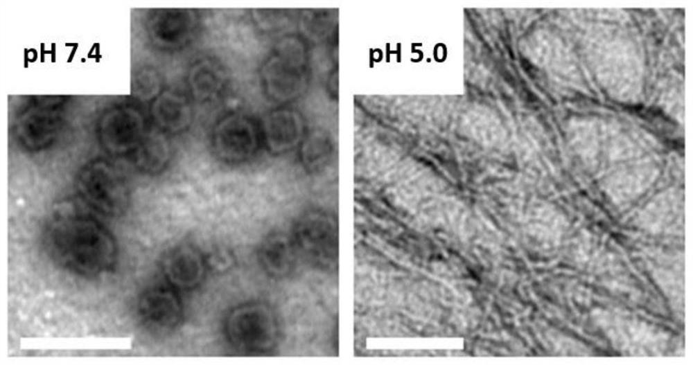
[0041] Dissolve 0.1mg of the polypeptide described in Example 1 in 1mL of ultrapure water, adjust the pH to 7.4 with 0.1M HCl solution and 0.1M NaOH solution, sonicate for 1 minute, and let stand for 30 minutes to prepare an electron microscope sample. Observe the self-assembled morphology, which appears to be a liposome-like structure (see figure 1 ).

[0042] Then the pH of the solution was adjusted to 5.0, and left to stand for 30 minutes to prepare an electron microscope sample, which was observed by a transmission electron microscope and showed a nanofiber morphology (see figure 1 ).

Embodiment 3

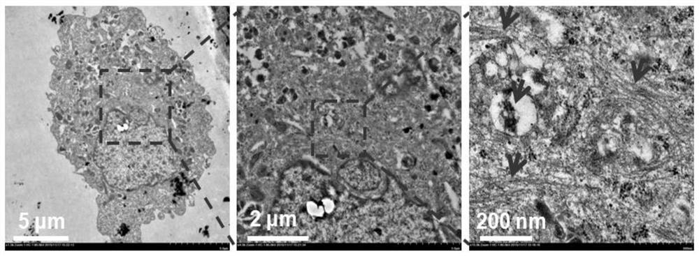
[0044] Observation of self-assembled nanofibers in cells by transmission electron microscope: Incubate the polypeptide with tumor cell MCF-7, collect the cells after 6 hours, centrifuge at 1000r / min at 4°C for 3 minutes, and remove the old medium on the upper layer. Then the cells were fixed with 2.5% glutaraldehyde and 1% osmic acid, dehydrated with different concentrations of ethanol, and embedded with epoxy resin. Then use Diamond Island to cut the cells into ultra-thin sections with a thickness of 60-90nm, place the sections on copper grids, stain with uranyl acetate and lead citrate, and observe the formation of intracellular fibers under a biological transmission electron microscope (HT7700) ( See figure 2 ).


PUM


Abstract

The invention relates to a self-assembled nanometer material, and specifically discloses a polypeptide and a polypeptide liposome formed by self-assembly thereof that can undergo shape transformation in tumor cell lysosomes. The main body of the polypeptide is a peptide chain formed by 6 alanines, the N-terminal of the peptide chain is modified with a group containing a benzene ring, and the C-terminal of the peptide chain is connected with an RGD polypeptide sequence. The polypeptide can self-assemble to form a liposome-like structure at a weakly alkaline physiological pH. After being recognized by tumor cells and entering the lysosome through endocytosis, the polypeptide responds to the acidic pH in the lysosome, and the self-assembled morphology Liposome structural transformation into nanofibers caused permeabilization of the lysosomal membrane, allowing more cathepsins to be released into the cytoplasm. When the polypeptide liposome is loaded with drugs capable of inhibiting lysosome repair or accelerating lysosome release of cathepsin, it can cooperate with polypeptide nanofibers to enhance cytotoxicity.

Description

technical field [0001] The present invention relates to self-assembled nanomaterials, in particular to a polypeptide self-assembled nanosystem with high tumor specificity and lysosome-targeted pH-responsive shape transformation. Background technique [0002] The process of molecular assembly is very common in nature. The coiling and folding of proteins and the double helix of DNA molecules are all biochemical processes under the control of self-assembly. Their delicate and mysterious forces even contain the mystery of the origin of life. The process of molecular self-assembly is a process that is not affected by external forces: it relies on molecular recognition between molecules or between a certain segment of a molecule and another segment, such as hydrogen bonds, van der Waals forces, electrostatic forces, hydrophobic interactions, Non-covalent weak interactions such as π-π stacking effects form molecular aggregates with a specific arrangement order. Once the process of ...

Claims


Application Information

Patent Timeline
Patent Type & Authority Patents(China)
IPC IPC(8): C07K19/00A61K9/127A61K47/42A61K45/00A61P35/00
CPCA61K9/127A61K45/00A61K47/42C07K7/06C07K2319/33
Inventor 赵颖聂广军覃好季天骄
Owner THE NAT CENT FOR NANOSCI & TECH NCNST OF CHINA
Why Eureka
Try Eureka