Prediction method of choroidal neovascularization based on constitutive model and finite element method

A new blood vessel and constitutive model technology, applied in 3D modeling, instrumentation, design optimization/simulation, etc., can solve the problem of not taking into account the different biological characteristics of different tissue structures, inaccurate prediction of large deformation changes, and only linear prediction Changes and other issues to achieve good prediction results and high accuracy

Active Publication Date: 2020-02-11
SUZHOU BIGVISION MEDICAL TECH CO LTD
View PDF6 Cites 1 Cited by
  • Summary
  • Abstract
  • Description
  • Claims
  • Application Information

AI Technical Summary

Problems solved by technology

Existing prediction methods for neovascularization growth usually only use a single growth model such as the finite element method for prediction. This method has certain shortcomings and limitations: ① It is based on the assumption of isotropy, ② It does not take into account the differences in different tissue structures. Biological characteristics, ③ can only predict linear changes, ④ can only predict changes in small deformations, inaccurate predictions of large deformation changes, etc.

Method used


Image

  • Prediction method of choroidal neovascularization based on constitutive model and finite element method
  • Prediction method of choroidal neovascularization based on constitutive model and finite element method
  • Prediction method of choroidal neovascularization based on constitutive model and finite element method

Examples


Embodiment Construction

[0055] Specific embodiments of the present invention are described in further detail below:

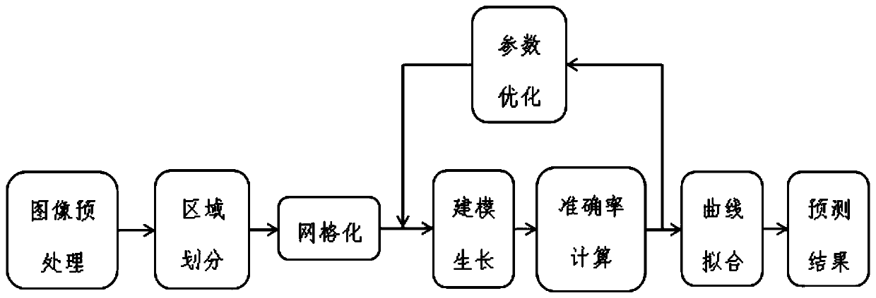
[0056] The basic block diagram of this method is as follows figure 1 As shown, it mainly includes the following steps:

[0057] 1) Image preprocessing

[0058] (a) Down-sampling processing: In order to improve processing efficiency and optimize memory usage, in this embodiment, the three-dimensional retinal SD-OCT image with 512*1024*128 pixels is down-sampled to 64*64*64 pixels.

[0059](b) OCT image registration was performed using CAVASS software (software developed by MIPG).

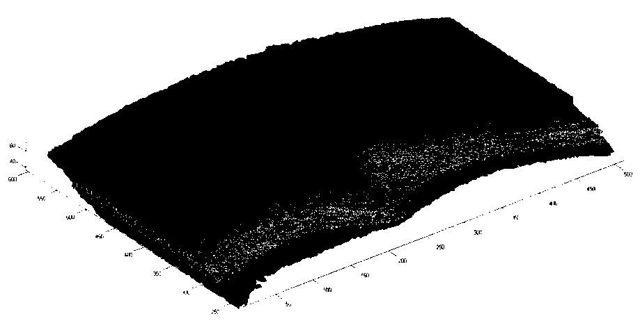
[0060] 2) Region extraction and division

[0061] Manually mark the gold standard to extract the retinal region of interest (CNV region) and divide the surrounding tissues (inner retinal layer, outer retinal layer, choroid layer);

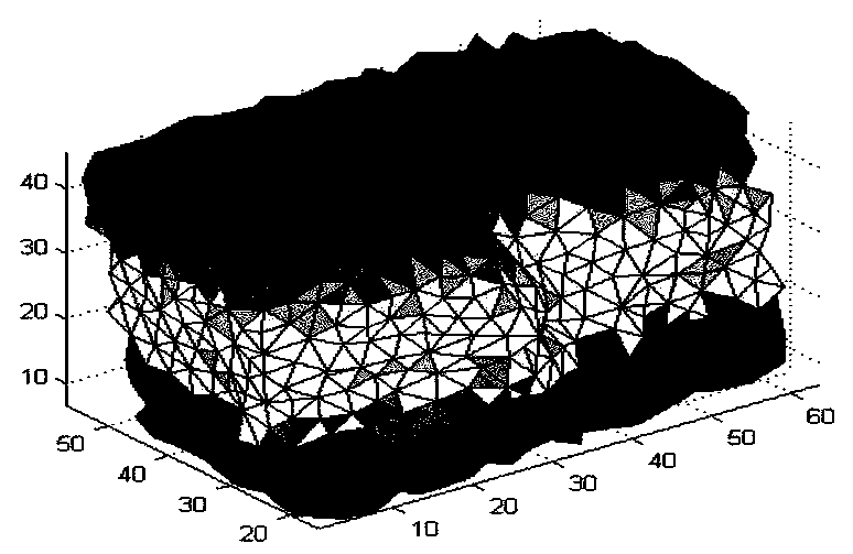
[0062] 3) Meshing, using a 3D surface and volumetric mesh generator for multiscale, adaptive tetrahedral mesh generation of the CNV region, inner retinal layer, outer retinal...


PUM


Abstract

The invention discloses a choroidal neovascularization growth protection method combining a constitutive model with a finite element. The method comprises the steps of image preprocessing; area division and partition, specifically including dividing an image into four areas consisting of a CNV area, an outer retina layer, an inner retina layer and a choroids layer; meshing, specifically including performing tetrahedral mesh generation on the four areas; modeling, specifically including modeling by using a hyperelastic biomechanical model and a reaction diffusion equation, and adding quality variation after choroidal neovascularization grows into an equation as a source item, thus causing a deformation gradient tensor to continuously change according to the growth of the new vessel; optimizing the model, computing the best accuracy rate, and performing parameter test; and fitting a parameter curve according to a parameter predicted at each time point, and predicting the growth parameter of the last time point to acquire a prediction result. According to the method provided by the invention, the biomechanical model can be built in a more flexible and personalized mode, the model assumes that organization is orthotropic, the good prediction results can be provided for non-linear large-deformation areas, and the accuracy is high.

Description

technical field [0001] The invention relates to the technical fields of computer vision, image processing and analysis, and belongs to a modeling method for growth prediction, in particular to a modeling method for choroidal neovascularization (CNV) growth prediction applied to SD-OCT (frequency-domain optical coherence tomography). Background technique [0002] The choroid is located between the retina and sclera and is composed of fibrous tissue, small blood vessels, and capillaries. The blood circulation of the choroid nourishes the outer layer of the retina, the lens, and the vitreous body. Due to the large blood flow and slow flow rate in the choroid, and its soft and thin characteristics, it is not tolerant to external influences or shocks, making it easy for pathogens to escape here. Retention can cause choroidal diseases and various fundus diseases. Therefore, studying the growth of choroidal neovascularization (CNV) can obtain a lot of important data, which can prov...

Claims


Application Information

Patent Timeline
Patent Type & Authority Patents(China)
IPC IPC(8): G06F30/23G06T17/00G06F119/14
CPCG06F30/23G06T17/00
Inventor 陈新建左畅朱伟芳
Owner SUZHOU BIGVISION MEDICAL TECH CO LTD
Why Eureka
Try Eureka