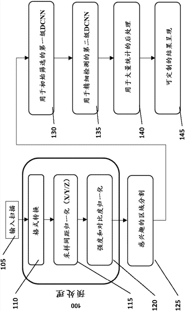
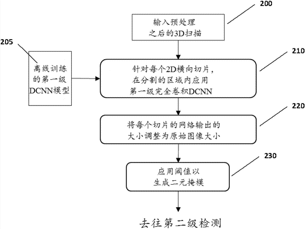
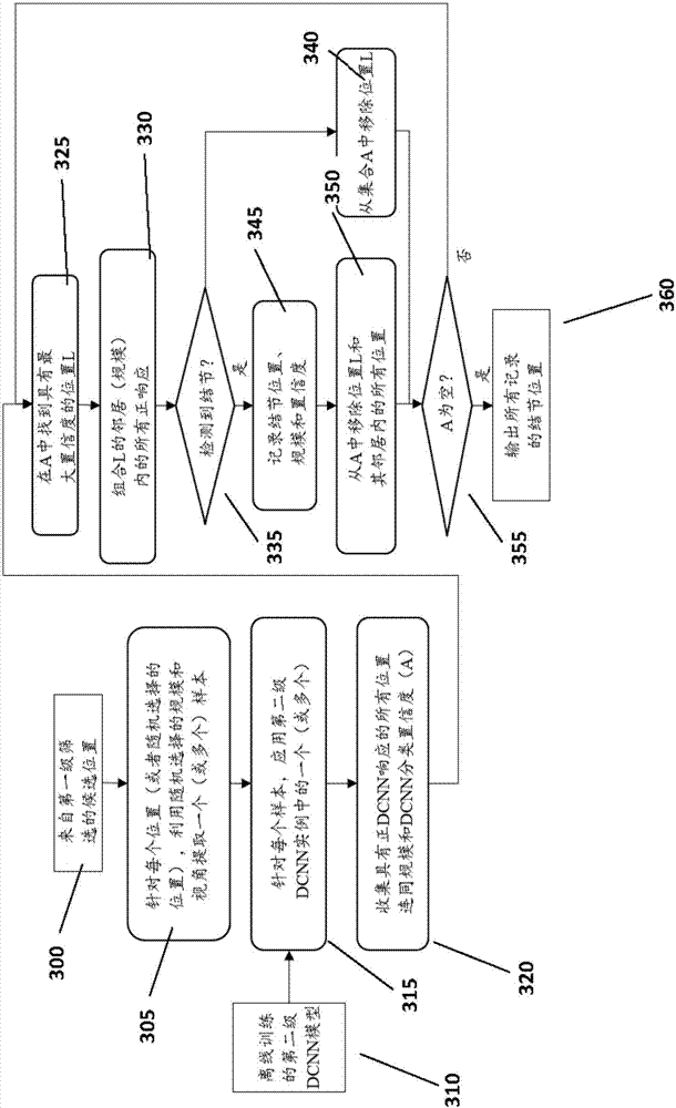
Computer-aided diagnosis system for medical images using deep convolutional neural networks
A convolutional neural network and medical image technology, which is applied in the field of computer-aided diagnosis system for medical images using deep convolutional neural network, can solve the problem of analyzing medical image length
- Summary
- Abstract
- Description
- Claims
- Application Information
AI Technical Summary
Problems solved by technology
Method used
Image
Examples
Embodiment 1
[0077] Example 1 - Report Generation
[0078] Figure 4 Examples of analysis inclusions are shown. The location of the nodule identified by the system is in the right upper lung. Nodules were determined to be non-solid and ground-glass-like. The x-y-z coordinates of the centroid of the nodule are 195-280-43. The diameter of the nodule is 11 mm along the long axis and 7 mm along the short axis. The borders of nodules are irregular in shape. The probability of malignancy is 84%.
[0079] Figure 5 An example of a diagnostic report is shown. In this embodiment, the report includes a user interface. The user is presented with three projected views 501 , 502 and 503 of a lung CT scan. Views 501, 502 and 503 include cursors to allow the user to pinpoint a location. Window 504 shows the 3D model of the segmented lung and detected nodule candidates (square points). The 3D model is superimposed in the projected views 501 , 502 and 503 .
Embodiment 2
[0080] Example 2 - Validation of pulmonary nodule detection
[0081] The techniques disclosed herein were applied to the publicly available lung nodule database (LIDC-IDRI) provided by the National Institutes of Health (NIH). The database included 1018 CT lung scans from 1012 patients. Scans were captured using various sets of CT machines and various parameter settings. Each voxel within the scan has been carefully annotated as normal (non-nodular) or abnormal (nodular) by four radiologists. In the training step, five-fold cross-validation is used for evaluation. All scans are first divided into five sets, each set containing approximately the same number of scans. Each scan was randomly assigned to one of five sets. Each evaluation iteration uses four of the five sets to train the lung nodule detector, and the remaining set is used to evaluate the trained detector. During training iterations, split and cascade detections are applied to the database. The evaluation was r...
PUM
 Abstract
Description
Claims
Application Information
 - R&D
- Intellectual Property
- Life Sciences
- Materials
- Tech Scout
- Unparalleled Data Quality
- Higher Quality Content
- 60% Fewer Hallucinations
Browse by: Latest US Patents, China's latest patents, Technical Efficacy Thesaurus, Application Domain, Technology Topic, Popular Technical Reports.
© 2025 PatSnap. All rights reserved.Legal|Privacy policy|Modern Slavery Act Transparency Statement|Sitemap|About US| Contact US: help@patsnap.com


