Wavelet-transform-based multi-modal medical image fusion method

A technology of medical image and fusion method, which is applied in the medical field to achieve the effect of improving reliability, strong robustness, and improving detail sensitivity

Active Publication Date: 2017-06-30
CARBON (SHENZHEN) MEDICAL DEVICE CO LTD
View PDF1 Cites 9 Cited by
  • Summary
  • Abstract
  • Description
  • Claims
  • Application Information

AI Technical Summary

Problems solved by technology

[0006] The purpose of the present invention is to propose a multimodal medical

Method used


Image

  • Wavelet-transform-based multi-modal medical image fusion method
  • Wavelet-transform-based multi-modal medical image fusion method
  • Wavelet-transform-based multi-modal medical image fusion method

Examples


Embodiment Construction

[0051] The present invention will be further described below in conjunction with the accompanying drawings and embodiments.

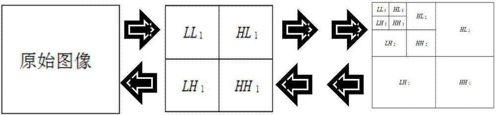
[0052] Such as Figure 1-4 As shown, a multimodal medical image fusion method based on adaptive wavelet base, which includes the following steps:

[0053] S1. Using an adaptive wavelet-based filter to perform wavelet transformation on medical images of different modalities, and decompose the aforementioned images into high-frequency, low-frequency, and high-low-frequency components;

[0054] S2. Superimpose the high-frequency, low-frequency, and high-low-frequency combined components obtained by decomposing any two medical images of different modalities to obtain the high-frequency, low-frequency, and high-low-frequency combined components of the fused image;

[0055] S3. Perform discrete wavelet inverse transform on the high-frequency, low-frequency and high-low-frequency combined components of the fused image to obtain a fused image of the original s...


PUM


Abstract

The invention relates to a multi-modal medical image fusion method based on an adaptive wavelet basis. The method comprises: S1, wavelet transform is carried out on medical images with different modes by using an adaptive wavelet basis filter, the images are decomposed into high-frequency, low-frequency and high-and-low-frequency-combined components respectively; S2, superposition of the high-frequency, low-frequency and high-and-low-frequency-combined components obtained by decomposition of any two different modal medical images is carried out to obtain high-frequency, low-frequency and high-and-low-frequency-combined components with images fused; and S3, discrete wavelet inverse transform is carried out on the high-frequency, low-frequency and high-and-low-frequency-combined components with images fused to obtain a fused image with the original size. Because of application of the wavelet transform technology to the image processing field, the detail sensitivity to low-frequency components by wavelet coefficients can be improved by the adaptive wavelet basis, so that the high-frequency and low-frequency components obtained by decomposition become accurate and thus the fusion effect can be improved.

Description

technical field [0001] The invention relates to the medical field, in particular to a method for fusing multimodal medical images using image processing technology, in particular to a multimodal medical image fusion method based on wavelet transform. Background technique [0002] At present, with the rapid development of computer science and technology and medical impact engineering, many advanced medical imaging devices have appeared in the world, providing medical images of various modalities for clinical medical diagnosis, and these images reflect the structure of the human body from different aspects , different information about organs and diseased tissues. [0003] For example, CT (Computed Tomography) images have strong spatial resolution and geometric characteristics, and the imaging of bones is very clear. It can provide a good reference for lesion location, but the contrast of soft tissues is relatively low. MR (Magnetic Resonance) images can clearly reflect the a...

Claims


Application Information

Patent Timeline
IPC IPC(8): G06T5/50
CPCG06T5/50G06T2207/10081G06T2207/20064
Inventor 王晓芳
Owner CARBON (SHENZHEN) MEDICAL DEVICE CO LTD
Why Eureka
Try Eureka