
An image processing method for simultaneously realizing CT perfusion and energy spectral liver scanning

A technology for image processing and perfusion scanning, which is applied in computer tomography scanners, instruments for radiological diagnosis, medical science, etc. Effects of sclerosis and cone beam artifacts, improving CT value accuracy, and increasing information for perfusion diagnosis

Active Publication Date: 2020-05-22
THE FIRST AFFILIATED HOSPITAL OF ZHENGZHOU UNIV
View PDF4 Cites 0 Cited by
  • Summary
  • Abstract
  • Description
  • Claims
  • Application Information

AI Technical Summary

Problems solved by technology

[0003] To sum up, the problems existing in the existing technology are: due to the limitations of CT machine hardware and software performance, the existing one-stop perfusion combined energy spectrum scanning technology can only achieve 40mm scanning, which is not conducive to providing more clinical diagnosis Information; the scanning time is basically fixed, and more perfusion information cannot be provided; the number of perfusion scanning layers is large, the scanning mode is basically fixed, and there are few parameters available for adjustment, which is not conducive to adjusting the radiation dose, and the scanning radiation dose is high; the perfusion breath-holding time is long , the image is greatly affected by respiratory artifacts; the operation is complicated and time-consuming, which is not conducive to clinical promotion

Method used


Image

  • An image processing method for simultaneously realizing CT perfusion and energy spectral liver scanning
  • An image processing method for simultaneously realizing CT perfusion and energy spectral liver scanning

Examples


Embodiment Construction

[0012] In order to make the object, technical solution and advantages of the present invention more clear, the present invention will be further described in detail below in conjunction with the examples. It should be understood that the specific embodiments described here are only used to explain the present invention, not to limit the present invention.

[0013] The application principle of the present invention will be described in detail below in conjunction with the accompanying drawings.

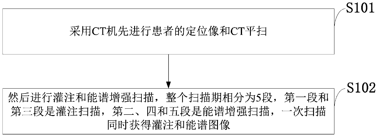
[0014] Such as figure 1 As shown, the image processing method for simultaneous perfusion and spectral liver scanning provided by the embodiment of the present invention includes the following steps:

[0015] S101: Use a CT machine to first perform a positioning image and a plain CT scan of the patient;

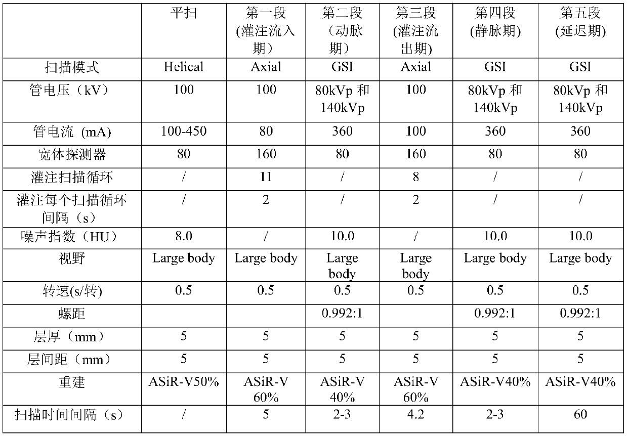
[0016] S102: Then perform a perfusion scan and an energy spectrum enhanced scan. The entire scan phase is divided into 5 sections. The first and third sections are the perfusion infl...


PUM


Abstract

The invention belongs to the technical field of perfusion and energy spectrum combined scanning, and discloses an image processing method capable of realizing CT perfusion and energy spectrum liver scanning simultaneously. According to the image processing method capable of realizing CT perfusion and energy spectrum liver scanning simultaneously, first, a CT machine is adopted for carrying out image locating and CT plain scanning on a patient, then, a contrast agent is injected, perfusion scanning and energy spectrum enhanced scanning are carried out, wherein the whole scanning period comprises 5 stages, the first stage and the third stage are perfusion scanning, the second stage, the fourth stage and the fifth stage are energy spectrum enhanced scanning, and through one-time scanning, perfusion and energy spectrum images are obtained simultaneously. The radiation dosage can be manually controlled and reduced, and therefore, the defects that in the existing similar technologies, the radiation dosage can not be regulated, and the radiation dosage is high are overcome; the perfusion image can provide the dynamic blood supply change characteristic of lesion, and the energy spectrum enhanced image can provide an image with the multiparameter mode beneficial for focus diagnosis and differential diagnosis; the perfusion image and the energy spectrum image can be obtained easily and conveniently through the one-time scanning method, thus abundant diagnosis information is provided for the clinic, and the radiation dosage of patients is reduced.

Description

technical field [0001] The invention belongs to the technical field of CT perfusion combined with energy spectrum scanning, and in particular relates to an image processing method for simultaneously realizing CT perfusion and energy spectrum liver scanning. Background technique [0002] The working principle of the existing one-stop CT perfusion combined with energy spectrum scanning technology is to scan the operating object according to the perfusion mode first, and then select any scanning phase to perform energy spectrum reconstruction according to the original image of perfusion through post-processing and reconstruction after scanning. Although this scanning technology can obtain perfusion images and energy spectrum images at the same time, this technology has the following disadvantages: 1) The range of energy spectrum reconstruction is limited to the range of perfusion scanning, and perfusion can only achieve 40mm scanning due to technical limitations , which is not ...

Claims


Application Information

Patent Timeline
Patent Type & Authority Patents(China)
IPC IPC(8): A61B6/03A61B6/00
CPCA61B6/032A61B6/481A61B6/50A61B6/507A61B6/5211
Inventor 吕培杰刘杰高剑波
Owner THE FIRST AFFILIATED HOSPITAL OF ZHENGZHOU UNIV
Learn More