Method and device for displaying internal marker of human organ three-dimensional medical model

A technology of human organs and marking points, which is applied in the field of image processing, can solve the problems of being unable to distinguish the inside or outside of the marking point, unable to effectively compare and view, and unable to distinguish the real position of the marking point, so as to achieve the effect of improving accuracy

Active Publication Date: 2017-09-22
QINGDAO HISENSE MEDICAL EQUIP
View PDF5 Cites 9 Cited by
  • Summary
  • Abstract
  • Description
  • Claims
  • Application Information

AI Technical Summary

Problems solved by technology

[0005] Due to the transparency of the 3D medical model of human organs, the visual positions of the marked points inside and on the surface of the 3D medical model of human organs are the same in the display results, and it is impossible

Method used


Image

  • Method and device for displaying internal marker of human organ three-dimensional medical model
  • Method and device for displaying internal marker of human organ three-dimensional medical model
  • Method and device for displaying internal marker of human organ three-dimensional medical model

Examples


Embodiment Construction

[0059] In order to make the object, technical solution and advantages of the present invention clearer, the implementation manner of the present invention will be further described in detail below in conjunction with the accompanying drawings.

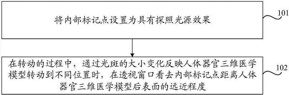
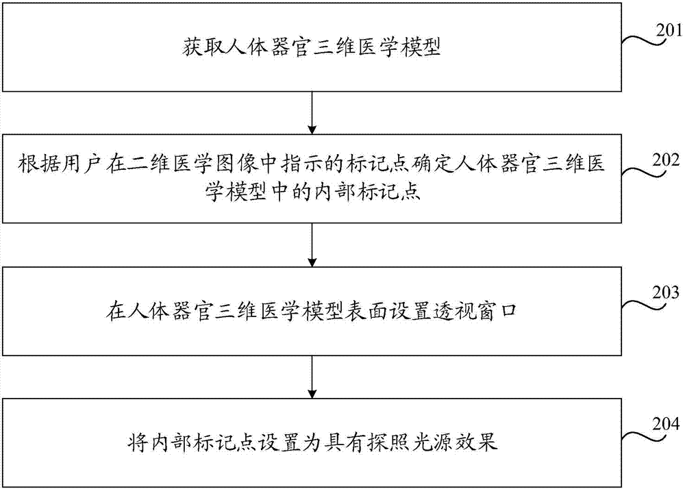
[0060] An embodiment of the present invention provides a method for displaying internal marker points of a three-dimensional medical model of human organs on a display screen. The internal marker point is a pre-marked point inside the three-dimensional medical model of human organs. Window, internal markers can be observed through the perspective window, when the three-dimensional medical model of human organs is manipulated to rotate, the position of the perspective window on the surface of the model also changes so that the internal markers can always be seen from the perspective window. Such as figure 1 As shown, the method may include:

[0061] Step 101. Set the internal marker points to have the effect of a searchlight source.

...


PUM


Abstract

The invention discloses a method and device for displaying an internal marker of a human organ three-dimensional medical model, which belongs to the field of image processing. A perspective window is arranged on the surface of the human organ three-dimensional medical model, so that the internal marker can be observed from the perspective window. When the model is manipulated to rotate, the position of the perspective window on the surface of the model also changes, so that the marker can always be seen from the perspective window. The marker is set to have the effect of a search light source, and the irradiation effect is that when the model is rotated, the irradiation direction is always back to the perspective window and the light source irradiates the rear surface of the human organ three-dimensional medical model to carry out reflection to form a light spot effect. In the process of rotation, the change in the size of the light spot can reflect the distance between the marker and the rear surface from the perspective window when the human organ three-dimensional medical model is rotated to different positions. According to the invention, the accuracy of the method for displaying the internal marker is improved, and the method and device are used for displaying the internal marker on a display screen.

Description

technical field [0001] The invention relates to the field of image processing, in particular to a method and device for displaying internal marker points of a three-dimensional medical model of human organs. Background technique [0002] With the development and application of medical imaging technologies such as computed tomography (English: Computed Tomography; abbreviation: CT), magnetic resonance imaging (English: Magnetic Resonance Imaging; abbreviation: MRI), ultrasound (English: Ultrasonography; abbreviation: US), traditional Two-dimensional images only express the anatomical information of a certain section, while three-dimensional visualization technology uses two-dimensional and spatial information to construct the relationship between the size and shape of human organs, soft tissues, lesions and surrounding tissues, which can assist doctors in diagnosis and treatment in a more vivid and three-dimensional manner. To improve the accuracy and efficiency of diagnosis ...

Claims


Application Information

Patent Timeline
IPC IPC(8): G06T17/00G06F19/00G06T15/80G06T19/00
CPCG06T15/80G06T17/00G06T19/003
Inventor 韩月乔田广野
Owner QINGDAO HISENSE MEDICAL EQUIP
Why Eureka
Try Eureka