Medical imaging method and medical imaging system
A medical imaging system and medical imaging technology, applied in the field of medical imaging methods and medical imaging systems, can solve problems affecting medical imaging accuracy, lack of system sensitivity, etc., to avoid loss of attenuation information, avoid excessive coverage, and improve accuracy. Effect
- Summary
- Abstract
- Description
- Claims
- Application Information
AI Technical Summary
Problems solved by technology
Method used
Image
Examples
Embodiment 1
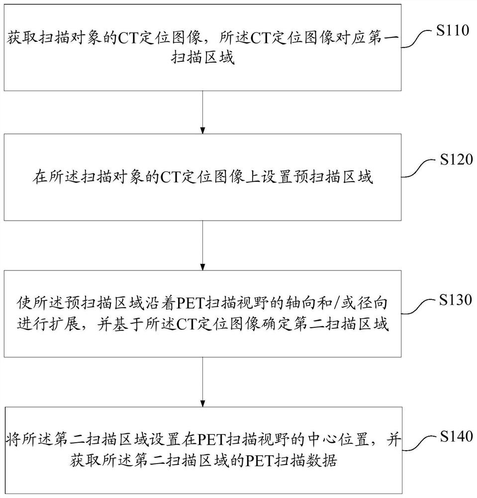
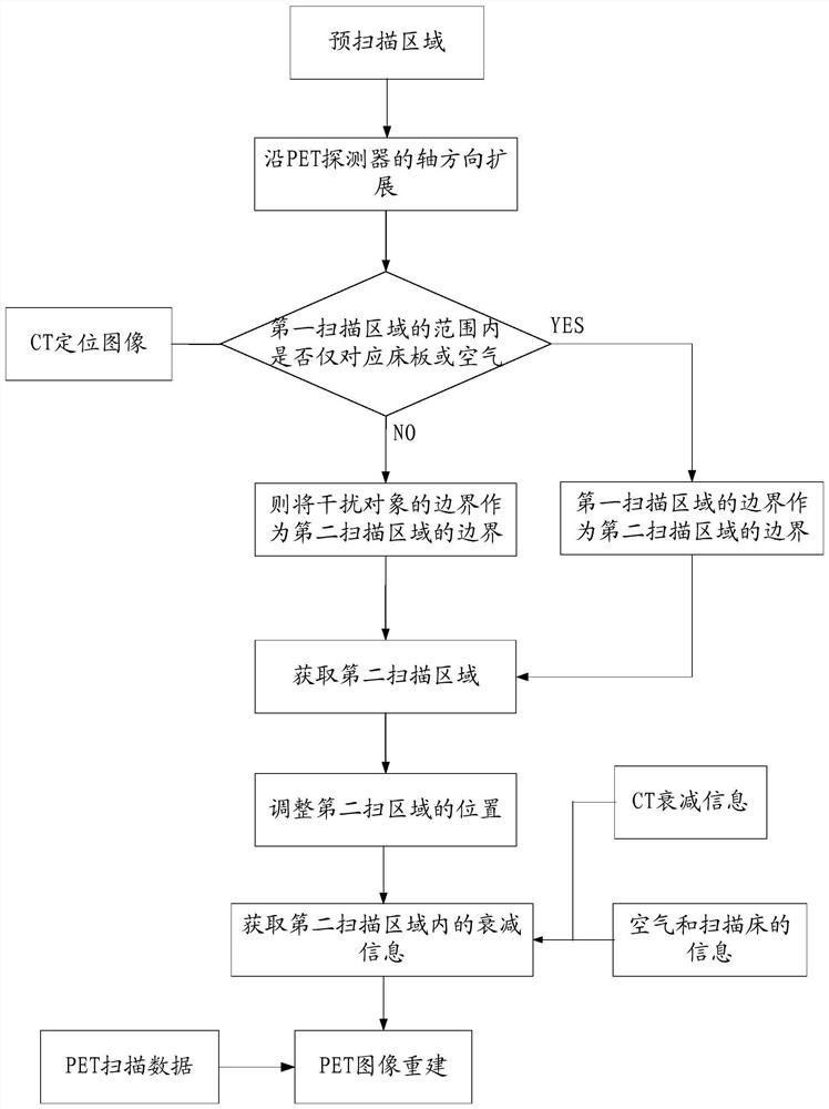
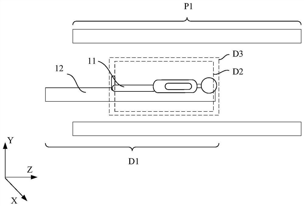
[0053] figure 1 It is a schematic flow chart of the medical imaging method in Embodiment 1 of the present invention, figure 2 It is a schematic diagram of the steps of the medical imaging method in Embodiment 1 of the present invention, Figure 3a and Figure 3b It is a schematic structural diagram during the process of determining the second scanning area in the medical imaging method in Embodiment 1 of the present invention. Combine below figure 1 , figure 2 and Figure 3a - Figure 3b As shown, the medical imaging method in this embodiment will be described in detail.
[0054] First, in step S110, a CT positioning image of the scanning object is acquired, and the CT positioning image corresponds to the first scanning area D1, that is, the first scanning area D1 is the scanning area of the CT positioning image. When subsequently determining the effective area for PET image reconstruction, the CT positioning image is used to determine the boundary of the effective ...
Embodiment 2
[0095] The difference from Embodiment 1 is that in this embodiment, interference objects other than the bed board and air are corresponding within the area range of the first scanning area D1. specific reference Figure 4a and Figure 4b Shown is a schematic structural diagram during the process of determining the second scanning region in the medical imaging method in Embodiment 2 of the present invention.
[0096] refer to Figure 4a As shown, the area range of the first scanning area includes the scanning object 11 , the bed board 12 , the air and the interfering object 14 , therefore, the first scanning area cannot be directly defined as the second scanning area. After the CT scan area D3 is defined, it is necessary to continue to expand the CT scan area D3, and obtain a second scan area based on the CT positioning image.
[0097] Specifically, continue to expand the area boundary toward the axis of the PET scanning field of view, for example:
[0098] First, expand to...
Embodiment 3
[0103] The invention also provides a medical imaging system. Figure 5 It is a schematic structural diagram of the medical imaging system in Embodiment 3 of the present invention, as Figure 5 As shown, the medical imaging system includes:
[0104] Bed board 110, used to carry the scanning object;
[0105] A CT scanning device 120, configured to acquire a CT positioning image of the scanned object;
[0106] A scan area defining unit 130, configured to set a pre-scan area on the CT positioning image, and expand the pre-scan area along the axial and / or radial direction of the PET scan field of view to determine a second scan area;
[0107] The PET detector 150 is configured to set the second scanning area at the center of the PET scanning field of view, and acquire PET scanning data of the second scanning area.
[0108] In addition, the medical imaging system further includes:
[0109] The attenuation information acquiring unit 140 is configured to acquire attenuation inform...
PUM
 Abstract
Description
Claims
Application Information
 - R&D
- Intellectual Property
- Life Sciences
- Materials
- Tech Scout
- Unparalleled Data Quality
- Higher Quality Content
- 60% Fewer Hallucinations
Browse by: Latest US Patents, China's latest patents, Technical Efficacy Thesaurus, Application Domain, Technology Topic, Popular Technical Reports.
© 2025 PatSnap. All rights reserved.Legal|Privacy policy|Modern Slavery Act Transparency Statement|Sitemap|About US| Contact US: help@patsnap.com


