Rational interpolation zooming method based on CT image edge

A technology of CT image and edge detection algorithm, applied in the field of image processing, can solve the problem that the image cannot correctly display the relationship between diseased tissue and normal tissue

Inactive Publication Date: 2017-11-07
SHANDONG UNIV OF FINANCE & ECONOMICS
View PDF3 Cites 11 Cited by
  • Summary
  • Abstract
  • Description
  • Claims
  • Application Information

AI Technical Summary

Problems solved by technology

In addition to hard defects such as medical hardware equipment and radiation dose limitation, the edge between lesion tissue and normal tissue in CT images is still relatively blurred. If

Method used


Image

  • Rational interpolation zooming method based on CT image edge
  • Rational interpolation zooming method based on CT image edge
  • Rational interpolation zooming method based on CT image edge

Examples


Embodiment Construction

[0043] The present invention will be further described below in conjunction with the accompanying drawings and embodiments.

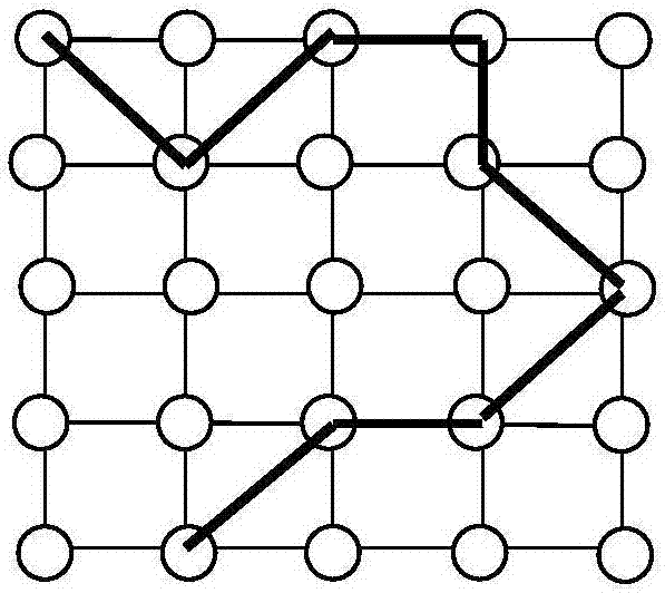
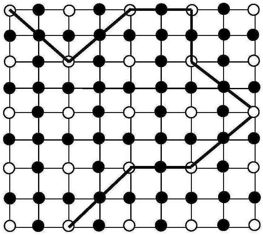
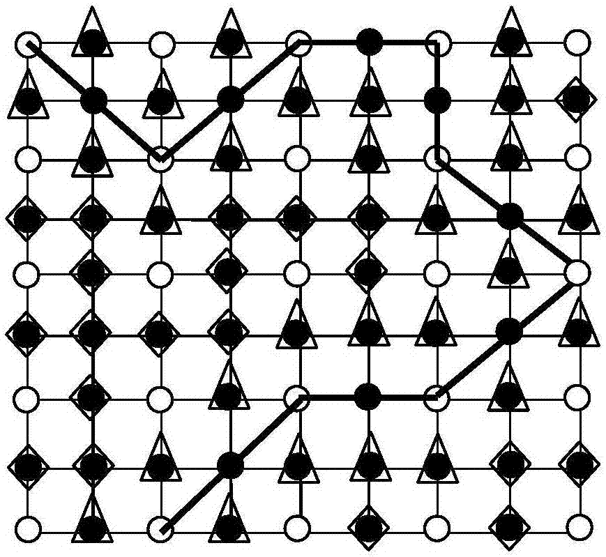
[0044] Aiming at the phenomenon that the boundary is easy to be blurred and distorted when the image is enlarged by using the existing method, the present invention discusses how to avoid losing the important factor of human vision—image edge information in the process of image zooming, so as to ensure the sharpness of the boundary contour after zooming in, and to maintain Clear image details and better subjective visual effects. To enlarge discrete images, one of the most effective methods is to construct the original surface of the image and perform high-density resampling. The invention proposes a CT image scaling method with good visual effect and maintaining edge features. The basic idea of ​​the present invention can be briefly described as follows: Generally, an image is composed of abrupt regions and smooth regions. The pixel values ​​in the s...


PUM


Abstract

The invention discloses a rational interpolation scaling method based on the edge of a CT image, which comprises step 1, obtaining the boundary of a single pixel of the image through an edge detection algorithm; step 2, obtaining a fitting curve of the boundary pixels, and resampling to obtain a new enlarged boundary; Step 3, resampling non-boundary pixels according to different situations. The boundary curve and key pixels are obtained through the edge detection algorithm, the fitting curve of the boundary pixels is obtained when zooming in, and the new boundary is obtained by resampling to maintain the sharpness of the boundary visually; non-boundary pixels are divided into rational interpolation and ordinary according to different situations There are two types of bilinear interpolation. Due to the special processing of the image edge, the loss of edge information during the image scaling process is avoided, so that the details of the image can be better displayed, and the image information of the lesion can be displayed better. The magnified CT image can clearly show the relationship between the lesion and the surrounding normal tissues and organs, so that some lesions that are not easy to be found can be detected early.

Description

technical field [0001] The invention relates to the technical field of image processing, in particular to a rational interpolation scaling method based on CT image edge features. Background technique [0002] Image scaling is a basic problem in image processing and has been widely used in practice. For example, image scaling is often used in medical, aerospace, and video applications. Its main purpose is to make the new image better reflect the original image. The shape of the scene, especially the detail information. In particular, in medical diagnosis, the observation of CT images, which is an important auxiliary diagnostic means for clinical disease diagnosis, often requires zoom-in operation so as to be suitable for details, especially the observation of lesions, so as to improve the accuracy of doctor's disease diagnosis. [0003] The essence of image magnification is to generate gray values ​​of unknown high-resolution pixels based on known gray values ​​of low-resolu...

Claims


Application Information

Patent Timeline
IPC IPC(8): G06T3/40G06T7/00G06T7/13G06T7/136
CPCG06T3/4007G06T7/0012G06T7/13G06T7/136G06T2207/10081G06T2207/20192G06T2207/30004
Inventor 高珊珊迟静兰婷婷王珊孟凡丽
Owner SHANDONG UNIV OF FINANCE & ECONOMICS
Why Eureka
Try Eureka