
An Image Detection Method to Improve Feature Matching Accuracy

A technology of image detection and feature matching, which is applied in the field of medical computers, can solve the problems affecting the real-time performance and long time of SIFT algorithm, and achieve real-time performance, improve matching accuracy, and improve the matching speed

Active Publication Date: 2021-02-02
BEIJING INSTITUTE OF TECHNOLOGYGY +1
View PDF3 Cites 0 Cited by
  • Summary
  • Abstract
  • Description
  • Claims
  • Application Information

AI Technical Summary

Problems solved by technology

It takes a long time for feature matching, which affects the real-time performance of the SIFT algorithm

Method used


Image

  • An Image Detection Method to Improve Feature Matching Accuracy
  • An Image Detection Method to Improve Feature Matching Accuracy
  • An Image Detection Method to Improve Feature Matching Accuracy

Examples


Embodiment Construction

[0043] The specific implementation process of the present invention will be described below in conjunction with the accompanying drawings.

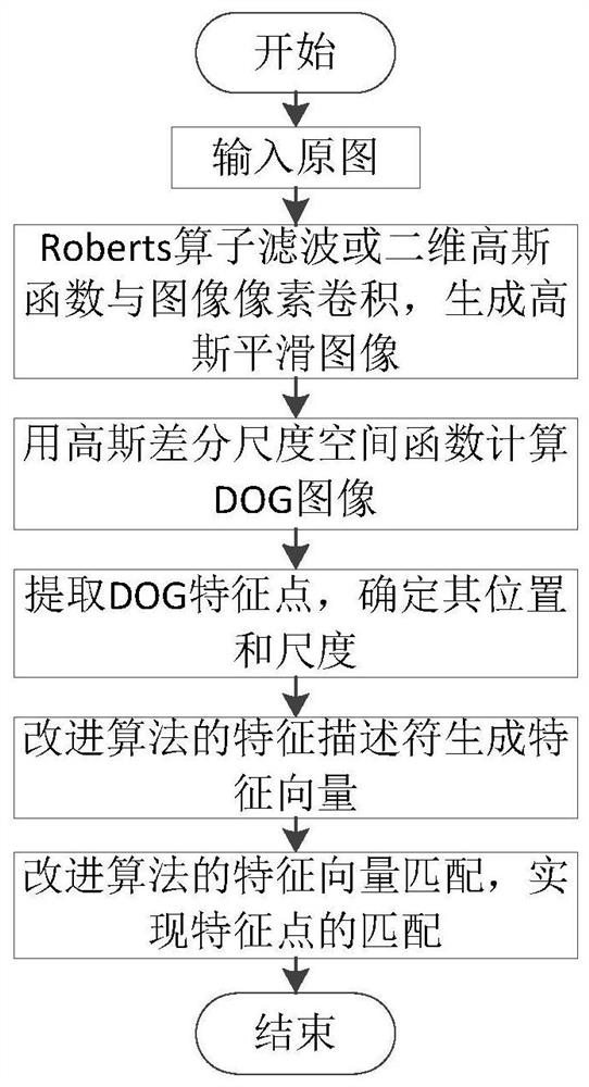
[0044] An image detection method that improves the accuracy of feature matching, the flow chart of the steps is as follows figure 1 shown. Specifically include the following steps.

[0045]Step 1. Use the Roberts operator to filter the image to be detected I(x,y) to generate a Gaussian smooth image; or: select a different scale factor σ, and combine the two-dimensional Gaussian function G(x,y,σ) with the image to be detected The pixels of I(x,y) are convolved to generate a Gaussian smooth image;

[0046] Step 2. Calculate the Gaussian smooth image output in the previous step by using the Gaussian difference scale space function to generate a DOG image;

[0047] Step 3, extracting the feature points of the DOG image, determining its position and scale;

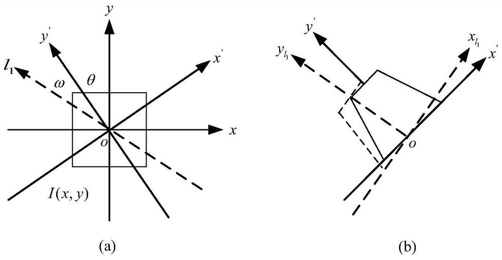
[0048] Step 4. Use Radon transformation to obtain a series of projection images on...


PUM


Abstract

The invention discloses an image detection method for improving feature matching precision, and belongs to the technical field of medical computers. The invention improves the SIFT algorithm, and uses the value obtained by Radon transformation of the image to form a feature vector. Using this method, the invention reduces it from 128 dimensions to 24 dimensions, thereby improving the real-time performance of the algorithm. At the same time, in order to improve the matching of feature vectors, the present invention selects two methods for removing false matches, first using structural similarity to roughly remove matching point pairs, and then using the distribution of spatial geometry and its Constraint conditions realize the fine elimination of matching points. On the basis of not affecting the image matching effect, the present invention can not only increase the matching speed, but also improve the matching accuracy and realize real-time performance.

Description

technical field [0001] The invention relates to an image detection method for improving feature matching accuracy, and belongs to the technical field of medical computers. Background technique [0002] After the 20th century, medical image technology has changed rapidly. Medical images can be divided into two types according to the information they provide. One is anatomical structure images, such as CT, MRI, B-ultrasound, etc., which have high pixel resolution and can display detailed anatomical information at a glance. The second is functional images, such as SPECT, PET, etc., which can completely display relevant information of organs, but the pixel resolution is relatively low, and some details of anatomy are powerless. Although these studies on medical images have been of great help, due to the limitations of providing image information, doctors need to combine their experience, spatial conception and speculation to judge the required information when making a diagnosi...

Claims


Application Information

Patent Timeline
Patent Type & Authority Patents(China)
IPC IPC(8): G06K9/62G06K9/44
CPCG06V10/34G06V10/751G06F18/22
Inventor 郭树理韩丽娜郝晓亭司全金林辉陈启明刘宏斌刘宏伟刘丝雨
Owner BEIJING INSTITUTE OF TECHNOLOGYGY
Learn More