
Mouse CT image kidney segmentation method based on random forest and statistic model

A CT image and random forest technology, applied in the field of medical image processing, can solve the problems of low accuracy and slow speed, and achieve the effect of improving the segmentation speed

Inactive Publication Date: 2017-12-22
NORTHWEST UNIV(CN)
View PDF4 Cites 9 Cited by
  • Summary
  • Abstract
  • Description
  • Claims
  • Application Information

AI Technical Summary

Problems solved by technology

[0003] To sum up, the problems existing in the prior art are: the current CT image organ segmentation method has low accuracy and slow speed.

Method used


Image

  • Mouse CT image kidney segmentation method based on random forest and statistic model
  • Mouse CT image kidney segmentation method based on random forest and statistic model
  • Mouse CT image kidney segmentation method based on random forest and statistic model

Examples


Embodiment Construction

[0045] In order to make the object, technical solution and advantages of the present invention more clear, the present invention will be further described in detail below in conjunction with the examples. It should be understood that the specific embodiments described here are only used to explain the present invention, not to limit the present invention.

[0046] The application principle of the present invention will be described in detail below in conjunction with the accompanying drawings.

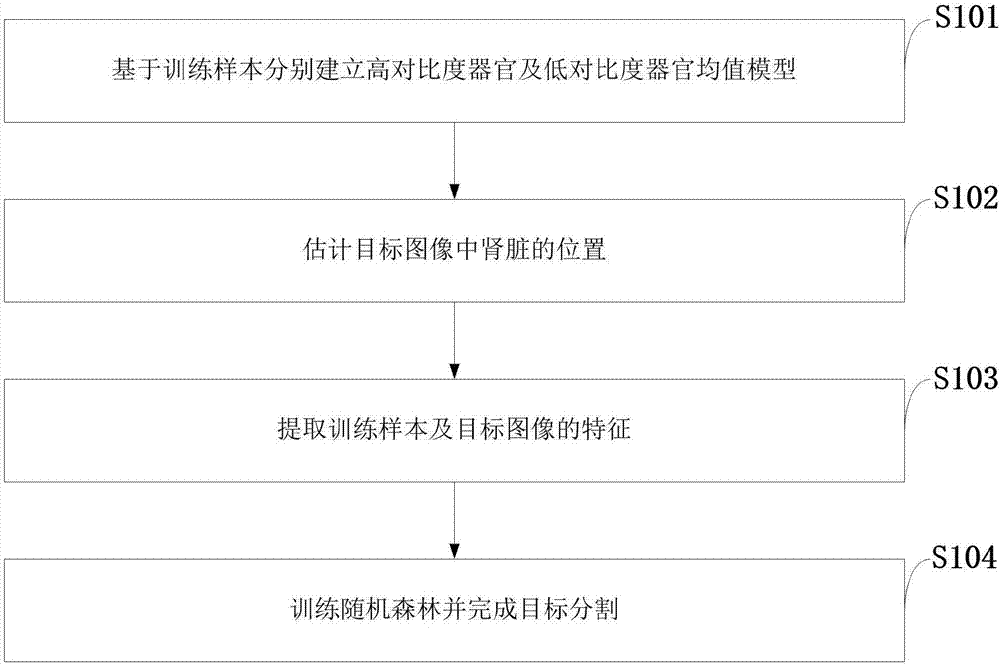
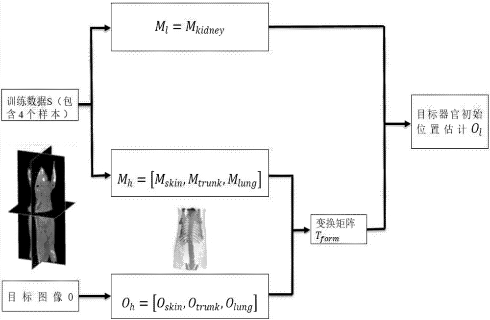
[0047] S101: Establishing high-contrast organ and low-contrast organ mean models based on the training samples;

[0048] S102: Estimate the position of the kidney in the target image;

[0049] S103: extracting features of training samples and target images;

[0050] S104: Train the random forest and complete target segmentation.

[0051] The application principle of the present invention will be further described below in conjunction with the accompanying drawings.

[0052] like f...


PUM


Abstract

The invention, which belongs to the technical field of medical image processing, discloses a mouse CT image kidney segmentation method based on a random forest and a statistic model. The method comprises: on the basis of a training sample, establishing a high-contrast-ratio organ mean value model and a low-contrast-ratio organ mean value model respectively; estimating the location of a kidney in a target image; extracting features of the training sample and the target image; and training a random forest and completing target segmentation. The feature expression for the CT image is constructed and thus the random forest can segment the CT image precisely, so tat problems of large data volume, complicated random forest calculation, and too low speed for the CT sequence image are solved. Meanwhile, an over-fitting problem caused by the statistic model is solved and a model can be established by using a few of samples; and kidney segmentation in the CT image is realized. The method having advantages of high precision, fast speed, and capability of being free of manual intervention has the great reference application value in fields like medical image segmentation.

Description

technical field [0001] The invention belongs to the technical field of medical image processing, and in particular relates to a mouse CT image kidney segmentation method based on a random forest and a statistical model. Background technique [0002] As an interdisciplinary subject with high practical value, medical imaging is receiving increasing attention, and its imaging technology is also constantly innovating, such as computed tomography (Computed Tomography, CT), ultrasound imaging (Ultrasonography, US) , Magnetic Resonance Imaging (MRI) and many other imaging techniques. As an important form of medical imaging, Micro-CT (micro computed tomography) has been widely used in clinical research on small animals. Medical images describe the detailed information of various organ tissues, structures and lesions, and provide an important basis for disease diagnosis, pathological location, anatomical structure research, surgical planning and guidance, etc. Due to the internal d...

Claims


Application Information

Patent Timeline
IPC IPC(8): G06T7/11G06T7/136
CPCG06T7/11G06T7/136G06T2207/10081G06T2207/30084
Inventor 侯榆青高培赵凤军贺小伟王宾易黄建曹欣
Owner NORTHWEST UNIV(CN)
Learn More