Spectral doppler detection

A technique of Doppler banding and Doppler estimation, applied in the direction of organ movement/change detection, blood flow measurement device, medical science, etc., can solve the problem of insufficient reduction of spots or other changes

Active Publication Date: 2018-01-09
SIEMENS MEDICAL SOLUTIONS USA INC
View PDF7 Cites 1 Cited by
  • Summary
  • Abstract
  • Description
  • Claims
  • Application Information

AI Technical Summary

Problems solved by technology

These passes use post-processing nonlinear filters, but may not sufficiently reduce speckle or other variations

Method used


Image

  • Spectral doppler detection
  • Spectral doppler detection
  • Spectral doppler detection

Examples


Embodiment Construction

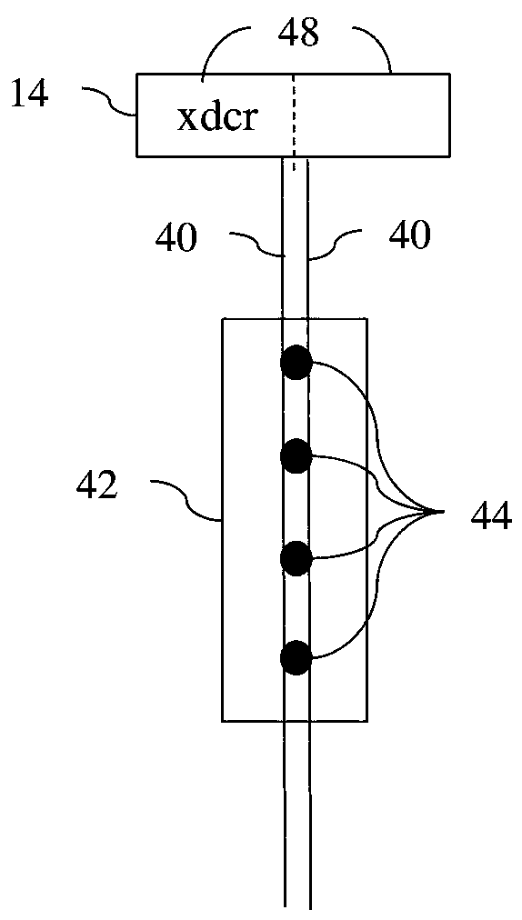
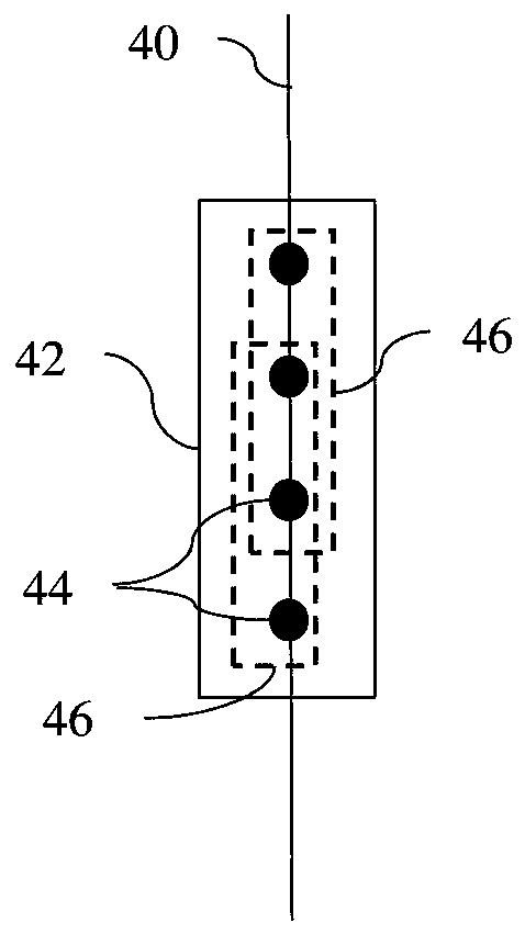
[0017] Interpretation of pulsed wave spectral imaging is often hampered by a "spotty" appearance of the spectrum and background noise, blurring the boundary between signal and noise. Pulsed wave Doppler detectability of the ultrasound spectrum can be improved. By creating samples that respond to different spatial components, multiple spectra can be estimated for a given Doppler gate, also with or without oversampling in time. Spectral variation is reduced by combining multiple spectra with at least partially uncorrelated noise. By reducing signal and noise variations, users can more easily distinguish flow from noise.

[0018] Multiple spectra are generated in a manner that does not require temporal oversampling, so the improvement in detectability is not limited by temporal sampling constraints. Multiple spectra are generated by: subdividing the sample volume into overlapping or non-overlapping sub-gates; using multiple receive per transmit with overlapping receive aperture...


PUM


Abstract

In spectral pulsed wave Doppler imaging, spatial variance in signal and / or noise are reduced by combination of multiple spectra with at least partially decorrelated noise. Rather than requiring oversampling in time, the multiple spectra for one Doppler gate are created from different spatial signals. The Doppler gate is divided into sub-gates, the beamformed sample locations in the Doppler gate are grouped into two or more groups using any selection criterion, and / or different receive apertures are used to simultaneously sample the Doppler gate. Spectra for the gate are estimated from the samples with the different spatial content and then combined.

Description

Background technique [0001] This embodiment relates to Doppler mode (eg spectral) imaging. By transmitting multiple pulses (eg, pulse waves (PW)) at a location, a Doppler response is generated. With spectral Doppler, the frequency spectrum of the motion or flow of an object for a single spatial region is estimated and displayed as a function of time. Spectral Doppler ultrasound imaging provides an image of the frequency spectrum of energy-modulated velocity values ​​(vertical axis) as a function of time (horizontal axis) for a gate. Spectra can be used to study fluid flow or tissue motion within a patient. [0002] Spectral Doppler in medical ultrasound is limited in sensitivity due to electrical and acoustic noise and signal processing dynamic range. In addition to using better quality electronic components and transducers, image processing can be used to reduce noise. For example, speckle noise is located in the Doppler stripes and is reduced. As another example, edges ...

Claims


Application Information

Patent Timeline
Patent Type & Authority Applications(China)
IPC IPC(8): A61B8/00
CPCA61B8/469A61B8/06A61B8/08A61B8/488A61B8/5269A61B8/543
Inventor 王景源P.D.弗赖布格尔
Owner SIEMENS MEDICAL SOLUTIONS USA INC
Why Eureka
Try Eureka