
Surgical operating microscope with integrated optical coherence tomography and display system

A technology for ophthalmic surgery and microscopy, applied in the field of surgical operating microscopes, can solve the problems of lack of OCT imaging, etc., and achieve the effect of simplifying surgical operations and reducing the number of adjustments

Inactive Publication Date: 2019-10-22
NOVARTIS AG
View PDF5 Cites 0 Cited by
  • Summary
  • Abstract
  • Description
  • Claims
  • Application Information

AI Technical Summary

Problems solved by technology

However, systems for providing real-time intraoperative OCT imaging to aid in visualization are still lacking

Method used


Image

  • Surgical operating microscope with integrated optical coherence tomography and display system
  • Surgical operating microscope with integrated optical coherence tomography and display system
  • Surgical operating microscope with integrated optical coherence tomography and display system

Examples


Embodiment Construction

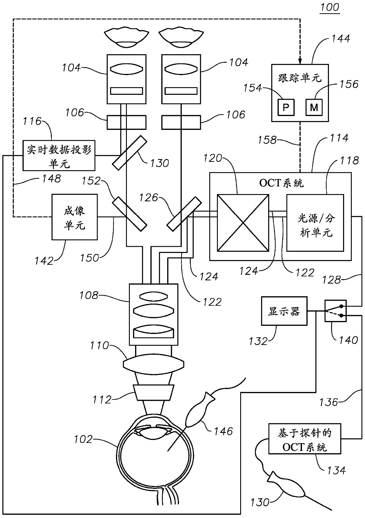
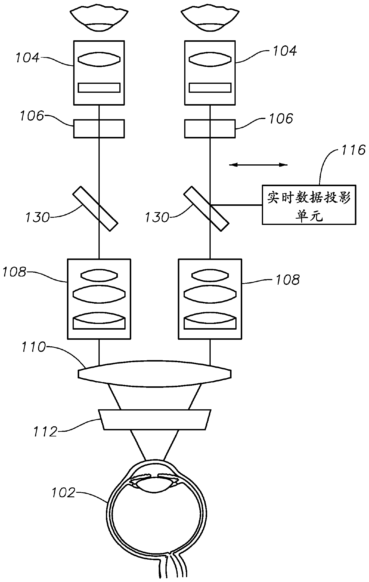
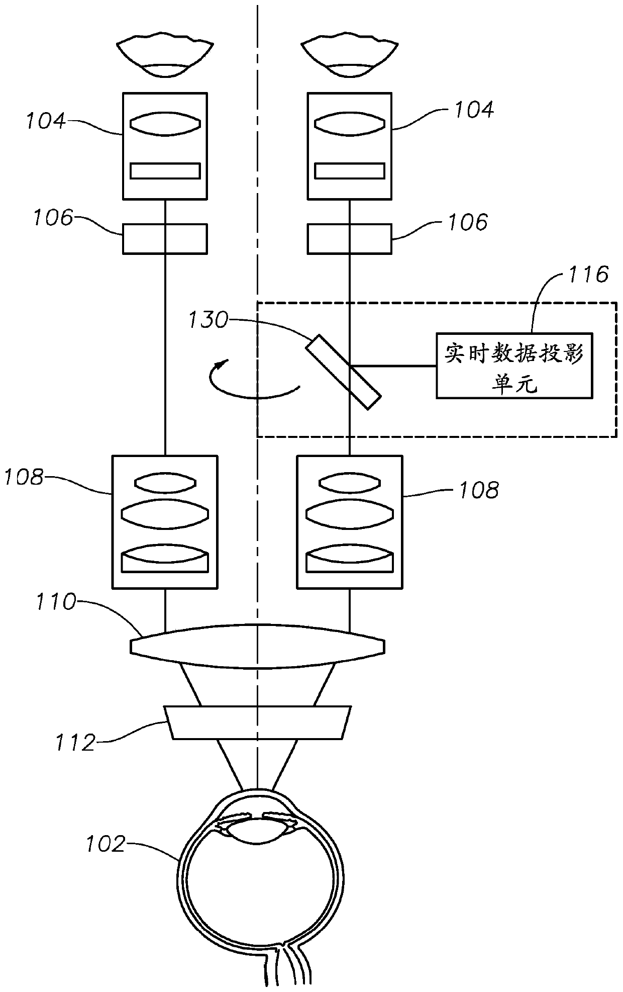
[0013] For the purposes of promoting an understanding of the principles of the disclosure, reference will now be made to the embodiments illustrated in the drawings, and specific language will be used to describe the same. However, it should be understood that no limitation of the scope of the present disclosure is intended. Any alterations and further modifications to the described systems, apparatuses, and methods, and any further applications of the principles of the disclosure will normally occur to those of ordinary skill in the art to which this disclosure pertains. In particular, it is entirely conceivable that the system, device and / or method described for one embodiment may be combined with the features, components and / or steps described for other embodiments of the present disclosure. However, for the sake of brevity, numerous iterations of these combinations will not be described individually. For simplicity, in some instances, the same reference numbers will be us...


PUM


Abstract

An ophthalmic surgery microscope comprising a beam coupler positioned between a first eyepiece and magnification / focus optics along an optical path of the surgical microscope, the beam coupler being operable to The OCT imaging beam is directed along a first portion of an optical path of the surgical microscope between the beam coupler and a patient's eye (the OCT image is generated based on a reflected portion of the OCT imaging beam). The surgical microscope further comprises: a real-time data projection unit operable to project OCT images produced by the OCT system; and a beam splitter along the surgical The optical path of the microscope is positioned between the second eyepiece and the magnification / focus optics. The beam splitter is operable to direct the projected OCT image along a second portion of the optical path of the surgical microscope between the beam splitter and the second eyepiece such that the projected OCT image can be viewed through the second eyepiece.

Description

technical field [0001] The present disclosure relates generally to surgical microscopes for ophthalmic surgery, and more particularly to surgical microscopes with integrated optical coherence tomography (OCT) and display systems. Background technique [0002] Eye surgeons often rely on surgical microscopes to see minute details of a patient's eye during eye surgery. One type of eye surgery - vitreoretinal surgery - involves vitrectomy (removal of the vitreous from the posterior chamber to gain access to the retina). Successful execution of vitrectomy requires essentially complete removal of the vitreous, including the most challenging region near the vitreous base. Due to the transparent nature of the vitreous, it can be challenging to perform a vitrectomy relying solely on a conventional surgical operating microscope for visualization. [0003] To aid in visualization, surgeons can rely on preoperative optical coherence tomography (OCT) imaging. OCT imaging is a method o...

Claims


Application Information

Patent Timeline
Patent Type & Authority Patents(China)
IPC IPC(8): A61B3/10A61B3/12A61B3/13
CPCA61B3/0041A61B3/102A61B3/13A61F9/007A61B90/361A61B90/37A61B2090/3735G02B21/0012G02B21/18G02B21/361A61B3/0025A61B3/10A61B3/12A61B3/132G02B13/04G02B13/16G02B15/24
Inventor 俞凌峰V·绍弗曼T·黑伦任虎刚
Owner NOVARTIS AG
Learn More