
A medical imaging scanning method and system

A scanning method and medical imaging technology, applied in the field of medical imaging scanning methods and systems, to achieve the effects of reducing scanning time, improving flexibility, and reducing scanning resource occupancy

Active Publication Date: 2021-06-29
SHANGHAI UNITED IMAGING HEALTHCARE
View PDF4 Cites 0 Cited by
  • Summary
  • Abstract
  • Description
  • Claims
  • Application Information

AI Technical Summary

Problems solved by technology

The supplementary scan method can solve the problem that the supplementary scan of the medical image sequence in the prior art can only be scanned backward from the end scan position

Method used


Image

  • A medical imaging scanning method and system
  • A medical imaging scanning method and system
  • A medical imaging scanning method and system

Examples


Embodiment Construction

[0034] In order to make the object, technical solution and advantages of the present invention clearer, the present invention will be further described in detail below in conjunction with the accompanying drawings. Obviously, the described embodiments are only some embodiments of the present invention, rather than all embodiments . Based on the embodiments of the present invention, all other embodiments obtained by persons of ordinary skill in the art without making creative efforts belong to the protection scope of the present invention.

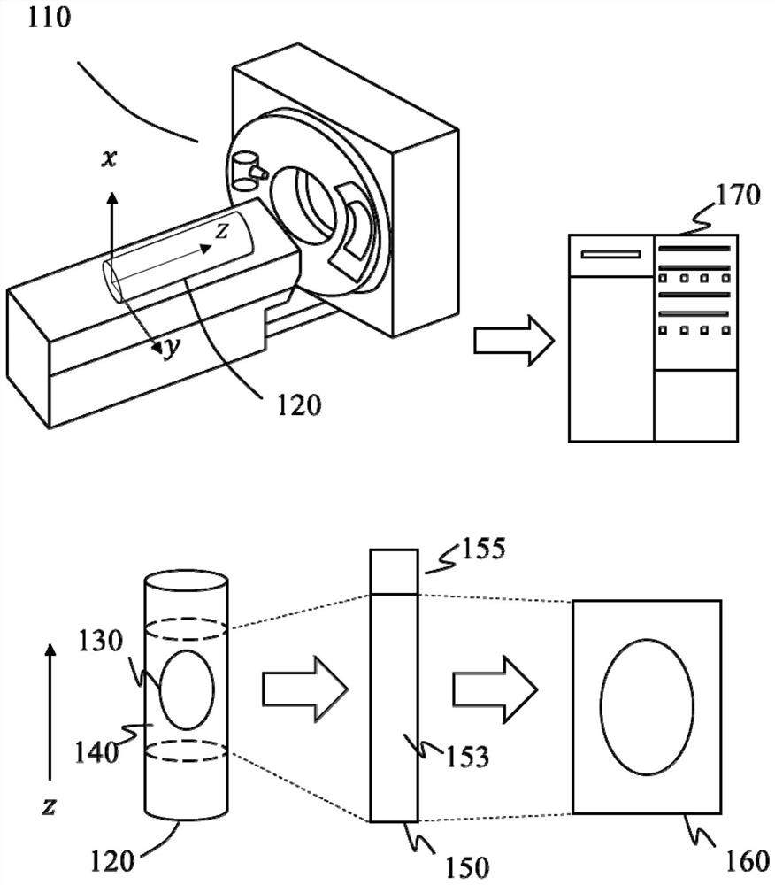
[0035] figure 1 It is a schematic diagram of a medical image sequence acquisition process provided according to an embodiment of the present invention. In order to obtain an image sequence 160 of a scanning target 130 in a scanning object 120 (for example, a human body, an animal body, etc.), the medical imaging device 110 can be used to scan The scan area 140 of the object 120 including the scan target 130 is scanned to generate scan data...


PUM


Abstract

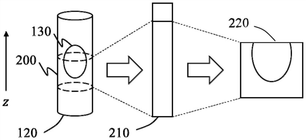
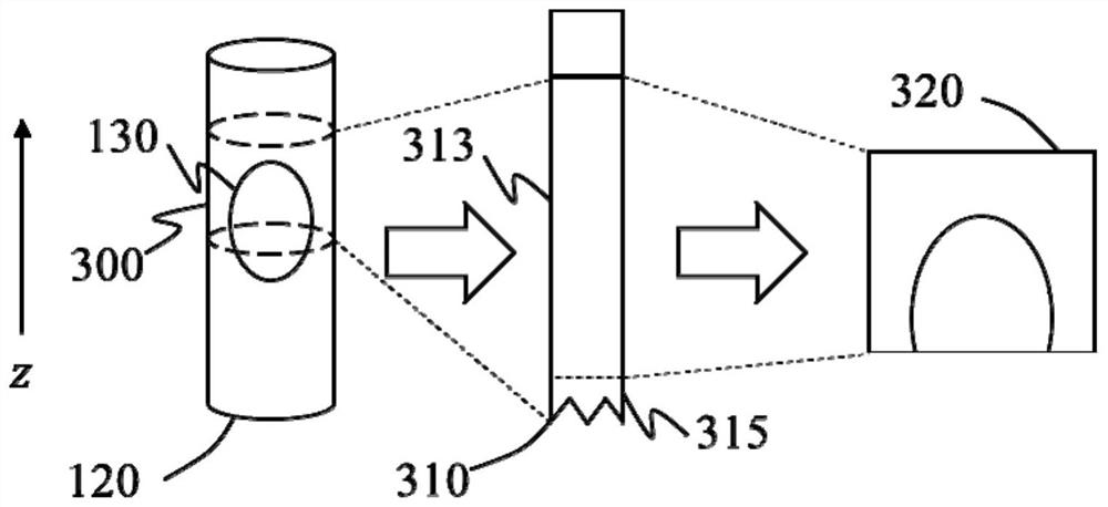
The invention discloses a medical imaging scanning method and a system for realizing the method. The method includes controlling the medical imaging device to perform a first scan on a first area of ​​the scan object to obtain first scan data, and determining a second area of ​​the scan object based on the first scan data, wherein the second area is connected to the first area or included in the first region. The method also includes controlling the medical imaging device to perform a second scan on a second region of the scan object to obtain second scan data, and generating a merged image sequence based on the first scan data and the second scan data, wherein the merged image sequence includes at least A sequence of images of part of the first area and at least part of the sequence of images of the second area. The invention can realize supplementary scanning from any position of the scanning object, and has the beneficial effects of improving the flexibility of supplementary scanning, reducing patient radiation dose absorption, reducing scanning time, and reducing scanning resource occupation.

Description

technical field [0001] The present invention relates to a medical imaging scanning method and system, in particular to a medical imaging supplementary scanning method and system. Background technique [0002] If there are deviations in the medical imaging scanning process (for example, missing scans, motion artifacts, unexpected interruption of scanning, etc.), it will often cause the medical images or image sequences generated based on this scan to be unusable (for example, for diagnosis, etc.) ). A medical image that can be used normally can be generated by performing a supplementary scan on the area where the scan has deviated, and merging the scan data and / or image sequence obtained from the supplementary scan with the scan data and / or image sequence obtained from the first scan sequence. Compared with re-scanning the entire area of ​​the target area, supplementary scanning can reduce the patient's absorption of radiation dose, reduce scanning time, and prevent the los...

Claims


Application Information

Patent Timeline
Patent Type & Authority Patents(China)
IPC IPC(8): G06T11/00
CPCG06T11/005G06T2207/30004
Inventor 王景波
Owner SHANGHAI UNITED IMAGING HEALTHCARE
Learn More