Self-positioning dental computed tomography scanning method and device

A tomography and computer technology, applied in the direction of dental radiology, diagnosis, clinical application of radiology, etc., can solve the problems of difficult popularization and installation, high device price, impact on human health, etc., and achieve the effect of avoiding unnecessary harm

Inactive Publication Date: 2018-04-06
慧友安控电子(深圳)有限公司
View PDF13 Cites 0 Cited by
  • Summary
  • Abstract
  • Description
  • Claims
  • Application Information

AI Technical Summary

Problems solved by technology

The use of head CT images for dental treatment has the following defects: 1. Doing the whole head CT because only the teeth need to be treated will have unnecessary impact on human health; It must be very clear; 3. The device is expensive and bulky, and it is not easy to popularize and install

Method used


Image

  • Self-positioning dental computed tomography scanning method and device
  • Self-positioning dental computed tomography scanning method and device
  • Self-positioning dental computed tomography scanning method and device

Examples


Embodiment Construction

[0035] The present invention will be further described below in conjunction with the accompanying drawings and embodiments.

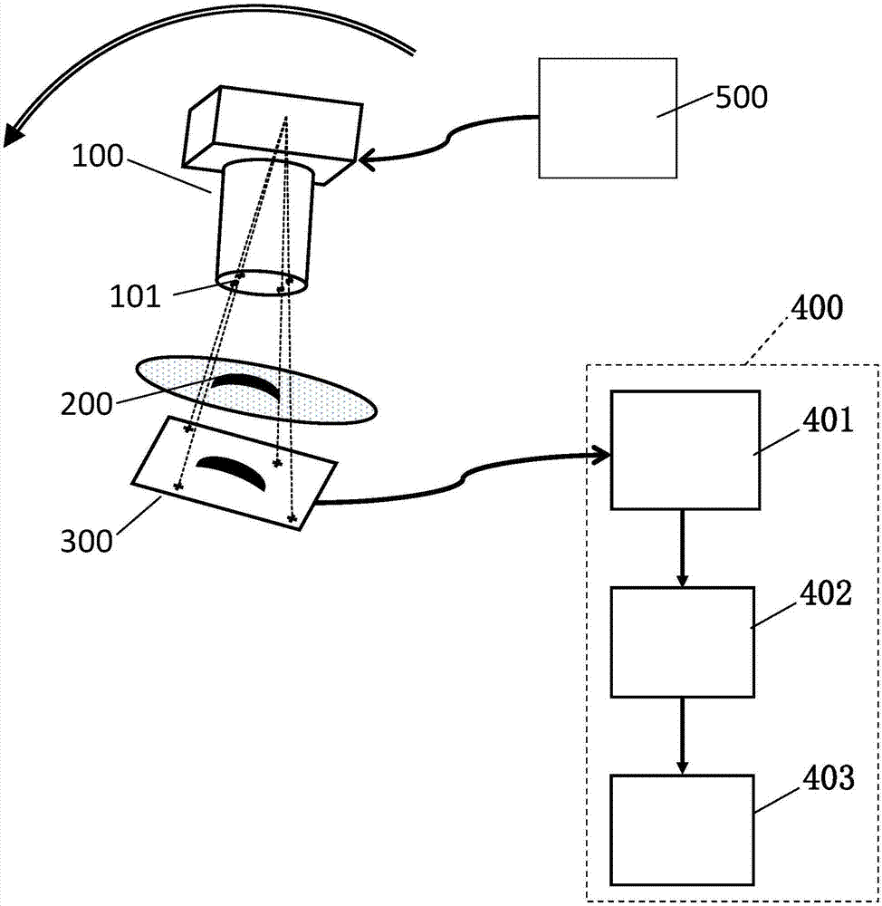
[0036] refer to figure 1 , some embodiments of the self-positioning tooth computed tomography device include: an X-ray image sensor 300 that can be placed on the inside of the tooth 200 to be tested; an X-ray source 100 that can be placed on the outside of the tooth 200 to be tested and rotate around the tooth 200; A rotation control device 500 that controls the rotation of the X-light source 100 ; a number of shading spots 101 arranged on the optical path of the X-light source 100 ; and a processing device 400 connected to the X-ray image sensor 300 . The processing device 400 includes: a punctuation recognition and positioning device 402 and a 3D graphics generation device 403 . The punctuation recognition and positioning device 402 is used to calculate the x-light source position and angle corresponding to the CT two-dimensional image; the 3D graphi...


PUM


Abstract

The invention provides a self-positioning dental computed tomography scanning method and device. The method includes the steps of placing an X-ray image sensor on the inner side of a to-be-measured tooth; setting an X-ray source on the outer side of the tooth; setting a plurality of light shielding mark points on an X-ray path; controlling the X-ray source to rotate around the tooth, and controlling the X-ray image sensor to acquire a plurality of CT 2D images during the rotation; calculating the X-ray source position and angle corresponding to each CT 2D image; and generating a 3D image of the tooth by using the CT 2D images and the corresponding X-ray source positions and angles. The scanning device includes the X-ray image sensor, the X-ray source, a rotation control device, the plurality of light shielding mark points, and a processing device connected to the X-ray image sensor. The method and device not only can obtain an accurate dental three-dimensional CT image, but also can avoid unnecessary harm to the human body caused by CT on the entire head.

Description

technical field [0001] The invention relates to a tooth CT (computerized tomography) image acquisition method and device, in particular to a self-positioning tooth computerized tomography method and device. Background technique [0002] Usually tooth CT is to place an X‐ray image sensor on the inside of the tooth, and fixedly set the X‐light source on the other side of the tooth, and the formed CT image is a planar image (two-dimensional image). Graphics are difficult to judge its position, which may cause bleeding and other problems during treatment. [0003] In head CT, the head is fixed in one position, and the X-light source rotates around the head to form a three-dimensional CT image. The use of head CT images for dental treatment has the following defects: 1. Doing the whole head CT because only the teeth need to be treated will have unnecessary impact on human health; It must be very clear; 3. The device is expensive and bulky, and it is not easy to popularize and i...

Claims


Application Information

Patent Timeline
Patent Type & Authority Applications(China)
IPC IPC(8): A61B6/14A61B6/00
CPCA61B6/14A61B6/466A61B6/5217
Inventor 黄佳铭
Owner 慧友安控电子(深圳)有限公司
Why Eureka
Try Eureka