Coronary artery image analysis method and data structure

A coronary artery and analysis method technology, applied in the field of image analysis, can solve the problems of increased difficulty in coronary angiography teaching, blurred boundaries of blood vessel walls, and difficulty in accurately defining boundaries.

Active Publication Date: 2018-04-20
ARMY MEDICAL UNIV
View PDF12 Cites 10 Cited by
  • Summary
  • Abstract
  • Description
  • Claims
  • Application Information

AI Technical Summary

Problems solved by technology

However, the biggest limitation of the traditional CTA-based imaging technology is that it can only clearly see the blood in the vessel lumen, that is, it can accurately obtain the boundary of the vessel lumen. The boundary of the vessel wall is relatively blurred, and it is difficult to accurately define its boundary. Therefore, it is quantified The analysis brings large errors, which makes the teaching of coronary angiography more difficult

Method used


Image

  • Coronary artery image analysis method and data structure
  • Coronary artery image analysis method and data structure
  • Coronary artery image analysis method and data structure

Examples


Embodiment 1

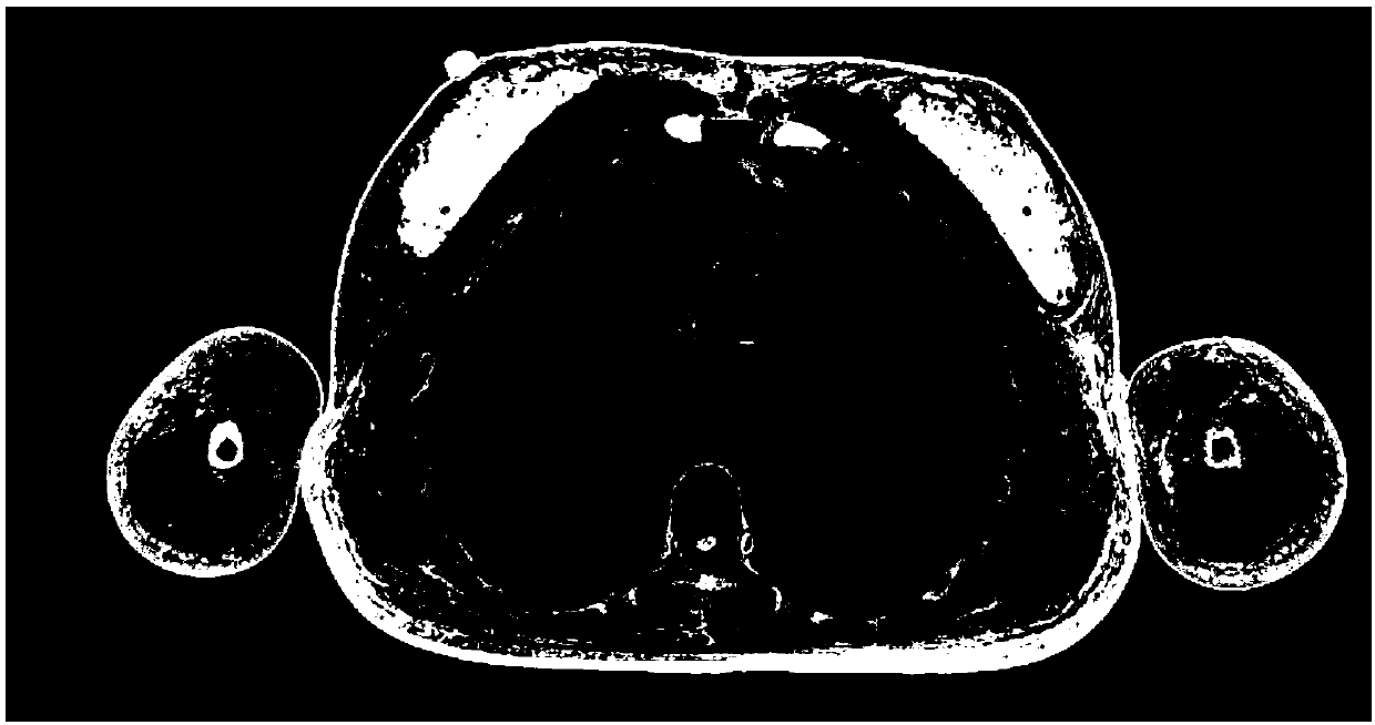
[0063] In this embodiment, the Chinese visualized human body data set is selected with reference to the human body data. The Chinese visualized human body data set is established by the Institute of Digital Medicine of the Army Military Medical University (formerly the Third Military Medical University). Human specimens without organic lesions are selected, and after shape measurement, The steps of perfusion, embedding and freezing were placed in the cryogenic laboratory and milled layer by layer with a TK-6350 CNC milling machine from head to toe, and were photographed layer by layer with a high-definition digital camera to obtain high-definition complete human body tomographic photos. Human body structure data set (Zhang Shaoxiang, Liu Zhengjin, etc. The first case of China's digital visual human body completed [J]. Journal of Third Military Medical University, 2002, (10): 24-10). The data set contains a total of 8 sets of human specimens, including data of different ages and...


PUM


Abstract

The invention provides a coronary artery image analysis method and a data structure. The coronary artery image analysis method comprises the steps of S1, performing preprocessing on reference human body data and detected human body data; S2, performing three-dimensional analysis and two-dimensional analysis on the reference human body preprocessed data and the detected human body preprocessed datato obtain reference human body macro and micro analysis data and detected human body macro and micro analysis data, and associating the reference human body analysis data and the detected human bodyanalysis data; and S3, synchronously displaying the reference human body analysis data and the detected human body analysis data. The beneficial effect is that users can simultaneously compare and observe two-dimensional image data, three-dimensional models and images in blood vessel cavities of coronary arteries of a reference human body and a detected human body so as to achieve better coronaryartery teaching effects.

Description

technical field [0001] The invention relates to an image analysis method, in particular to a coronary artery image analysis method and a data structure. Background technique: [0002] CT coronary angiography, also known as CTA (Computed Tomography Angiography, computed tomography angiography), refers to injecting a contrast agent into the blood vessel to make the blood vessel visualized under CT. The development process can be photographed to see the changes in the geometric shape of blood vessels more clearly. [0003] The analysis of coronary angiography images can play a role in the teaching process in the field of coronary medicine. However, the biggest limitation of the traditional CTA-based imaging technology is that it can only clearly see the blood in the vessel lumen, that is, it can accurately obtain the boundary of the vessel lumen. The boundary of the vessel wall is relatively blurred, and it is difficult to accurately define its boundary. Therefore, it is quant...

Claims


Application Information

Patent Timeline
Patent Type & Authority Applications(China)
IPC IPC(8): G06T7/00G06T7/11G06T7/136G06T11/00
CPCG06T7/0014G06T7/11G06T7/136G06T11/003G06T2207/10081G06T2207/10024G06T2207/30101G06T2207/30048G06T2211/404
Inventor 李颖刘丽
Owner ARMY MEDICAL UNIV
Why Eureka
Try Eureka