
Medical support air bag device and use method thereof

A technology of airbags and air chambers, applied in medical science, other medical devices, dilators, etc., can solve the problems of narrow treatment and operation space, and achieve the effect of easy operation and simple structure

Pending Publication Date: 2018-07-10
SHANDONG PROVINCIAL HOSPITAL
View PDF6 Cites 5 Cited by
  • Summary
  • Abstract
  • Description
  • Claims
  • Application Information

AI Technical Summary

Problems solved by technology

[0004] The purpose of the present invention is to provide a medical support air bag device and its usage method, which can solve the problem of narrow operation space in endoscopic treatment of upper esophageal lesions

Method used


Image

  • Medical support air bag device and use method thereof
  • Medical support air bag device and use method thereof
  • Medical support air bag device and use method thereof

Examples


Embodiment 1

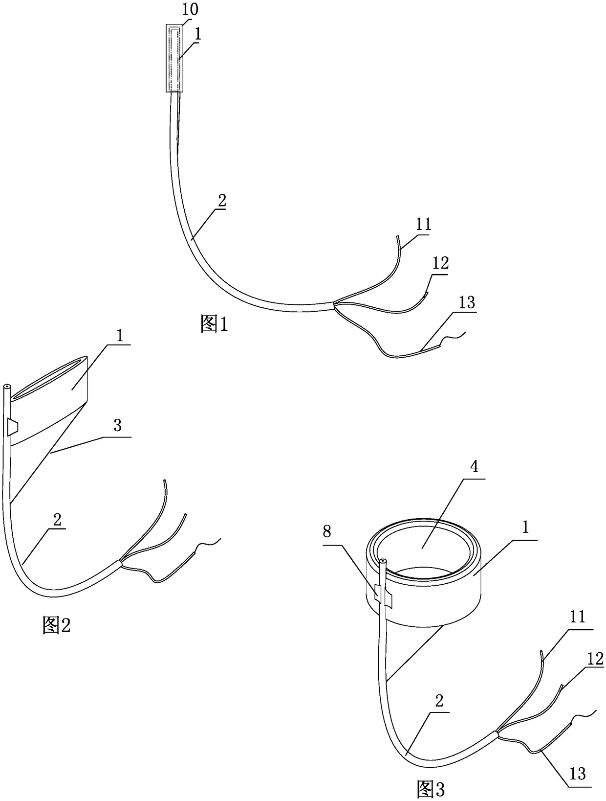
[0035] like figure 1 and figure 2 As shown, a medical support balloon device is mainly used to support the esophagus during esophageal endoscopic treatment to form a sufficient operating space, including a balloon 1, a three-lumen tube 2, a guide wire, an adjustment wire 3 and a balloon protection Sheath 10.

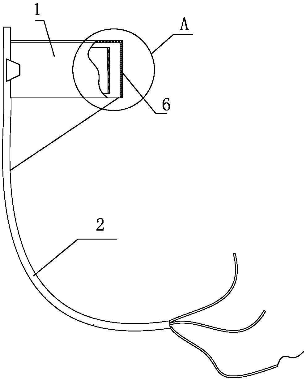
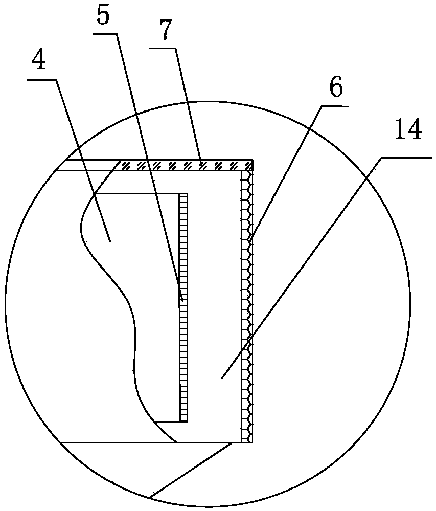
[0036] like figure 2 As shown, the airbag 1 adopts a hollow cylindrical cavity structure, similar to a swimming ring, and is a dry vacuum airbag when it is not inflated; image 3 As shown, when inflated, the airbag 1 bulges and can play a supporting role under the action of air pressure. like image 3 , Figure 4 and Figure 5 As shown, the airbag 1 includes a hollow cavity 4, an inner membrane 5 close to the hollow cavity 4, an outer membrane 6 far away from the hollow cavity 4, and an air cavity 14 between the inner membrane 5 and the outer membrane 6. The hollow Cavity 4 is used to pass through the endoscope when the balloon 1 enters the esophagus; the inner ...

Embodiment 2

[0043] like Figure 8 As shown, the front end of the three-lumen tube 2 runs through one side of the airbag 1 , and the joint between the airbag 1 and the three-lumen tube 2 is tightly pressed together. The ventilation lumen 11 of the three-lumen tube communicates with the air cavity 14 of the airbag 1 .

Embodiment 3

[0045] like Figure 9 As shown, further, the rear end of the ventilation lumen 11 is provided with an inflation valve 15, and the inflation valve 15 is used to connect with the front end of the syringe when inflated; the ventilation lumen 11 at the front end of the inflation valve 15 is provided with Air valve 16 is arranged. The air valve 16 includes a valve housing 17, an inner plug body 18 and a rotating rod 19. Preferably, the valve housing 17 adopts a circular cavity structure, and the front and rear ends of the valve housing 17 are respectively sealed with the ventilation lumen 11. Connected, one side of the valve casing 17 is provided with a rotating through hole; the inner plug body 18 adopts a circular structure, the inner plug body 18 is installed in the cavity of the valve casing 17, and the outer wall of the inner plug body 18 is close to the The inner wall of the valve housing 17 is provided with a vent hole 20 on the inner plug body 18, and the inner plug body 18 ...


PUM


Abstract

The invention aims at providing a medical support air bag device and a use method of the medical support air bag device, for solving the problem that the operation space is narrow during the treatmentprocess adopting the gastrointestinal endoscopy for the upper esophageal lesion. The medical support air bag device comprises an air bag, a three-cavity tube and an adjusting line, wherein the air bag adopts an annular air bag structure and is mounted at the front end of the three-cavity tube; the inside of the three-cavity tube comprises an inflating tube cavity, a guide tube cavity and an adjusting tube cavity, and the front end of the inflating tube cavity communicates with an air cavity of the air bag; the guide tube cavity used for allowing a guide wire to pass through penetrates throughthe front and rear ends of the three-cavity tube; the adjusting line is positioned in the adjusting tube cavity, the front end of the adjusting line is connected with the air bag, and the rear end ofthe adjusting line extends out of the rear end of the three-cavity tube. The medical support air bag device and the use method have the beneficial effects that the device can support the large treatment operation space under the endoscopy, so that the implementation of the treatment is facilitated.

Description

technical field [0001] The invention relates to the technical field of medical airbag devices, in particular to a medical support airbag device and its usage method. Background technique [0002] In the technical field of esophageal digestive endoscopy and endoscopic treatment, for upper esophageal lesions, including early cancer of the upper esophagus, benign submucosal tumors, and some other diseases that require endoscopic treatment, when performing endoscopic treatment, due to the location of the lesion It is relatively close to the entrance of the esophagus, and the esophagus has a natural physiological stenosis, resulting in a lack of operating space for endoscopic treatment, which makes such patients lose the opportunity for endoscopic treatment. [0003] The present invention combines the above problems encountered in the clinical treatment process and the Shandong Provincial Hospital "Selection and Comparison of Endoscopic Treatment Methods for Muscularis Propria Tu...

Claims


Application Information

Patent Timeline
IPC IPC(8): A61B17/00A61M29/04
CPCA61M29/02A61B17/00234A61B2017/00238A61B2017/00292A61B2017/0034A61M2210/105
Inventor 许昌芹许洪伟
Owner SHANDONG PROVINCIAL HOSPITAL
Learn More