Medical device for removing an implanted object using laser cut hypotubes

A technology of objects and blood vessels, applied in the field of devices for separating tissues, can solve the problems of increasing the risk and difficulty of removing lead wires

Active Publication Date: 2018-07-31
SPECTRANETICS
View PDF16 Cites 7 Cited by
  • Summary
  • Abstract
  • Description
  • Claims
  • Application Information

AI Technical Summary

Problems solved by technology

Tissue growth along the SVC and at other locations along the innominate vein may increase the risk and difficulty of l...

Method used


Image

  • Medical device for removing an implanted object using laser cut hypotubes
  • Medical device for removing an implanted object using laser cut hypotubes
  • Medical device for removing an implanted object using laser cut hypotubes

Examples


Embodiment Construction

[0160] Before any embodiments of the disclosure are explained in detail, it is to be understood that the disclosure is not limited in its application to the details of construction and the arrangement of parts set forth in the following description or shown in the following drawings. The disclosure is capable of other embodiments and of being practiced or operated in various ways. Also, it is to be understood that the phraseology and terminology used herein are for the purpose of description and should not be regarded as limiting. The use of "comprising", "comprising" or "having" and variations thereof herein is meant to encompass the items listed thereafter and equivalents thereof as well as additional items.

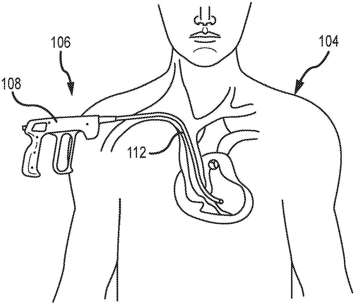
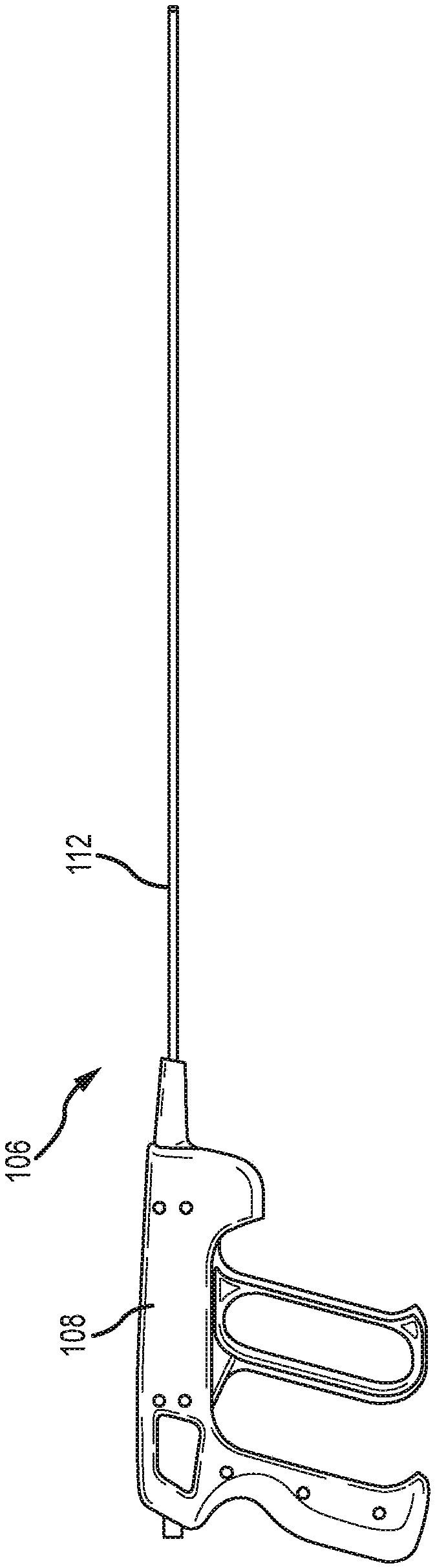
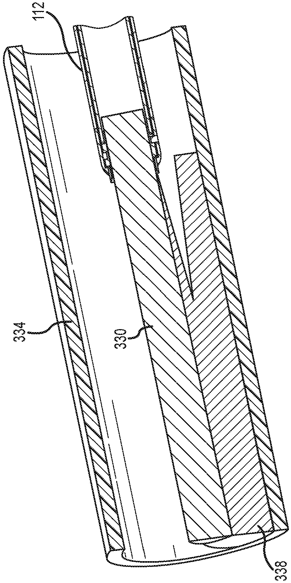
[0161] Embodiments according to the present disclosure provide a surgical device that includes a sheath assembly that can be safely deployed within a patient's vasculature and connects an implanted object, such as a lead, to a The patient's vasculature is isolated. f...


PUM


Abstract

Methods and devices for separating an implanted object, such as a pacemaker lead, from tissue surrounding such object in a patient's vasculature system. Specifically, the surgical device includes a handle, an elongate inner sheath and a circular cutting blade that extends from the distal end of the sheath upon actuating the handle. The circular cutting blade is configured to engage the tissue surrounding an implanted lead and cut such tissue in a coring fashion as the surgical device translates along the length of the lead, thereby allowing the lead, as well as any tissue remaining attached tothe lead, to enter the device's elongate shaft. The surgical device has a barrel cam cylinder in the handle assembly that imparts rotation of the blade and a separate cam mechanism in the tip of outer sheath assembly that imparts and controls the extension and retraction of the blade. The barrel cam cylinder and cam mechanism cooperate to cause the blade to rotate in a first direction and extendfrom and retract in the outer sheath due to a first actuation of the handle and to rotate in a second direction and extend and retract in the outer sheath due to a second actuation of the handle. Theinner sheath and outer sheath are constructed of laser-cut hypotubes, thereby allowing the surgical device, particularly the sheath assembly, to have a smaller profile for navigating smaller sized vasculature.

Description

[0001] Cross References to Related Applications [0002] This application is a continuation-in-part of U.S. Application No. 14 / 627,851, filed February 20, 2015, and entitled "MEDICAL DEVICE FOR REMOVING AN IMPLANTED OBJECT," which claims 35 BENEFITS AND PRIORITIES OF U.S.C. §119 AND / OR 35 U.S.C. §120, FILLED MARCH 13, 2014 AND TITLE "SURGICAL INSTRUMENT FORREMOVING AN IMPLANTED OBJECT" continuation-in-part of International Application PCT / US2014 / 026496, which claims the U.S. Benefit and priority of Provisional Application No. 61 / 793,597 under 35 U.S.C. § 119(e). U.S. Application No. 14 / 627,851, filed February 20, 2015 and entitled "MEDICAL DEVICE FOR REMOVING AN IMPLANTED OBJECT," also requires the following application to be filed at 35 U.S.C. § 119( BENEFITS AND PRIORITIES UNDER e): U.S. PROVISIONAL APPLICATION No. 61 / 947,377, FILING MARCH 3, 2014, TITLE "MEDICAL DEVICE FOR REMOVING AN IMPLANTED OBJECT"; 2014 U.S. Provisional Application No. 62 / 058,790, filed October 2, 201...

Claims


Application Information

Patent Timeline
IPC IPC(8): A61N1/05A61B17/3205A61B17/32
CPCA61B17/320016A61B17/32002A61B17/32053A61B2017/00309A61B2017/320032A61N2001/0578A61B17/3468A61N1/0573
Inventor K·P·格拉斯W·H·李B·E·卡加里斯R·L·卡弗
Owner SPECTRANETICS
Why Eureka
Try Eureka