Automatic lung medical image segmentation method

An automatic segmentation and medical image technology, applied in image analysis, image enhancement, image data processing, etc., can solve the problems of not giving the outline of the lungs, inaccuracy, etc.

Inactive Publication Date: 2018-08-28
HEBEI NORTH UNIV
View PDF5 Cites 8 Cited by
  • Summary
  • Abstract
  • Description
  • Claims
  • Application Information

AI Technical Summary

Problems solved by technology

[0003] To sum up, the problems existing in the existing technology are: the existing segmentation algorithms for lung lesion tissue based on region growth all need to manually obtain the initial growth ...

Method used


Image

  • Automatic lung medical image segmentation method
  • Automatic lung medical image segmentation method
  • Automatic lung medical image segmentation method

Examples


Embodiment Construction

[0046] In order to make the object, technical solution and advantages of the present invention more clear, the present invention will be further described in detail below in conjunction with the examples. It should be understood that the specific embodiments described here are only used to explain the present invention, not to limit the present invention.

[0047] The application principle of the present invention will be further described below in conjunction with the accompanying drawings.

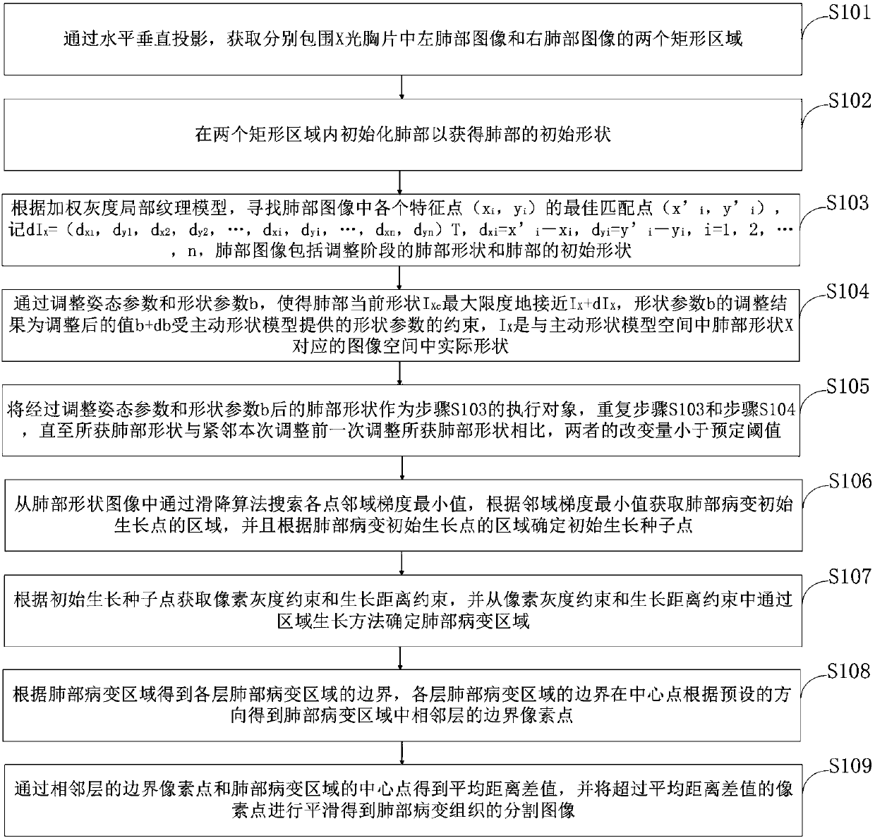
[0048] like figure 1 As shown, the present invention provides a lung medical image automatic segmentation method comprising the following steps:

[0049] Step S101, through horizontal and vertical projection, obtain two rectangular areas respectively surrounding the left lung image and the right lung image in the X-ray chest film;

[0050] Step S102, initialize the lungs in two rectangular areas to obtain the initial shape of the lungs;

[0051] Step S103, according to the weighted gr...


PUM


Abstract

The invention belongs to the medical image segmentation technology field and discloses an automatic lung medical image segmentation method. According to the method, the pixel gray constraint and the growth distance constraint are acquired according to an initial growth seed point, and a lung lesion region is determined from the pixel gray constraint and the growth distance constraint, and the lunglesion region is smoothed to accurately acquire a segmented image of the lung lesion tissue. The method is advantaged in that the initial lung shape can be relatively excellently acquired, over-segmentation during subsequent adjustment (for example, recognizing the spine and the stomach cavity as the lung region) can be avoided, under constraints of an active shape model, the lung region on the X-ray chest can be accurately segmented to provide valuable data for clinical or computer analysis.

Description

technical field [0001] The invention belongs to the technical field of medical image segmentation, in particular to an automatic segmentation method for lung medical images. Background technique [0002] At present, lung segmentation can be roughly divided into two methods: rule-based reasoning and pixel classification. The methods used by rule-based schemes are (local) thresholding, region growing, edge detection, morphological operations, fitting geometric models and functions, dynamic programming, etc., and most lung region segmentation algorithms fall into this category. Pixel-based classification schemes attempt to classify each pixel in an image into an anatomical type (usually lungs and background, but in some cases more types are used, e.g. also heart, middle diaphragm and diaphragm). The classifiers used are various neural networks or Markov random field models, and various (local) features are used for classification, including grayscale, position, and texture me...

Claims


Application Information

Patent Timeline
IPC IPC(8): G06T7/11G06T7/187
CPCG06T2207/10116G06T2207/30061G06T7/11G06T7/187
Inventor 梁俊花赵志升宋宸晏李静刘洋张晓彭一
Owner HEBEI NORTH UNIV
Why Eureka
Try Eureka