A pneumoconiosis detection method and system based on deep learning

A technology of deep learning and detection methods, applied in image analysis, image enhancement, instruments, etc., can solve problems such as complicated method process, great energy spent by doctors, strong subjective dependence of diagnostic results, etc., to reduce medical resources and reduce waiting time Effect

Active Publication Date: 2021-08-03
四川元匠科技有限公司
View PDF7 Cites 0 Cited by
  • Summary
  • Abstract
  • Description
  • Claims
  • Application Information

AI Technical Summary

Problems solved by technology

[0003] Since the diagnosis and differentiation of pneumoconiosis requires doctors to observe from multiple angles and for a long time, it has the following defects: (1) The patient cannot get the exact diagnosis result immediately, which affects life and work and subsequent treatment; (2) The traditional method process It is complicated and requires a lot of energy from doctors; (3) The diagnostic results are highly subjective, and different doctors may have different diagnostic results

Method used


Image

  • A pneumoconiosis detection method and system based on deep learning
  • A pneumoconiosis detection method and system based on deep learning
  • A pneumoconiosis detection method and system based on deep learning

Examples


Embodiment 1

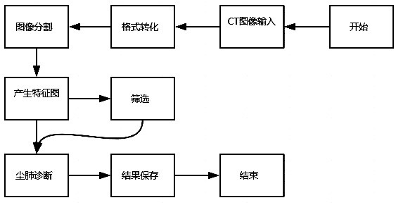
[0038] Such as figure 1 As shown, a deep learning-based pneumoconiosis nodule detection method provided in Embodiment 1 includes the following steps:

[0039] Step S1, the CT machine scans a complete chest image of the patient, starts the image conversion program, converts the CT image in DICOM format into a lung image in numpy array format, and reads the CT image data information.

[0040] In step 1, the CT image information refers to the patient information in the DICOM format data, the length and width of the CT image, and the interval between image pixels. Each dimension of the numpy array is n, h, w. n represents the channel of the image, h represents the height of the image, and w represents the width of the image.

[0041] Step S2, performing morphological operations to obtain a parenchymal image in which only the lung parenchyma is preserved.

[0042] In step 2, the specific operation steps of the morphological operation are as follows:

[0043] S2.1. Using a thres...

Embodiment 2

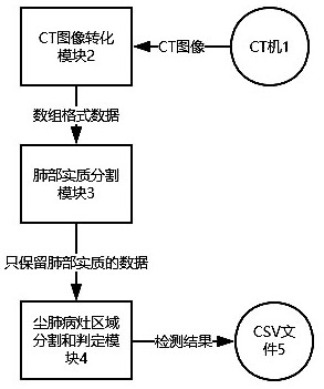
[0059] like figure 2 As shown, a deep learning-based pneumoconiosis nodule detection system provided in Embodiment 2 includes a CT machine 1, a CT image conversion module 2, a lung parenchyma segmentation module 3, a pneumoconiosis lesion area segmentation and determination module 4, and detection results The saving module (the csv file of the data saved by the detection result saving module) 5, the CT image conversion module 2, the lung parenchyma segmentation module 3, the pneumoconiosis lesion area segmentation and judgment module are programmed in Python language; the CT image scanned by the CT machine is input into the CT image The conversion module 2, the input of the lung parenchyma segmentation module 3 is the output of the CT image conversion module 2, the input of the pneumoconiosis lesion area segmentation and judgment module 4 is the output of the lung parenchyma segmentation module 3, and the detection result preservation module 5 is saved in the database The dat...


PUM


Abstract

The invention relates to a detection method for pneumoconiosis nodules, comprising the following steps: S1, converting a CT image in DICOM format into a lung image in numpy array format and reading the CT image data information; S2, performing morphological operations to obtain only retained The real image of the lung parenchyma; S3, dividing the parenchymal image into several small block images; S4, sending these small block images into the convolutional neural network for screening and diagnosis of pneumoconiosis nodules, and obtaining the detection results; S5. Saving the detection result in a csv format. The invention realizes automatic diagnosis of pneumoconiosis symptoms by converting, morphologically operating, and screening CT images to detect the number of pneumoconiosis nodules and the coordinate area where they are located, and reduces the problem that a large number of medical resources are required for pneumoconiosis identification.

Description

technical field [0001] The invention relates to a pneumoconiosis detection technology for CT images, in particular to a pneumoconiosis nodule detection technology based on deep learning. Background technique [0002] With the increasing population and demand for pulmonary medical care, there is an urgent need to speed up the speed and quality of nodule detection in pulmonary medical care. Among them, the problem of diagnosis and differentiation of pneumoconiosis is particularly prominent. The traditional differential diagnosis of pneumoconiosis requires lung biopsy of the patient and reference to the patient's previous X-ray chest films and case summaries. [0003] Since the diagnosis and differentiation of pneumoconiosis requires doctors to observe from multiple angles and for a long time, it has the following defects: (1) The patient cannot get the exact diagnosis result immediately, which affects life and work and subsequent treatment; (2) The traditional method process ...

Claims


Application Information

Patent Timeline
Patent Type & Authority Patents(China)
IPC IPC(8): G06T7/00G06T7/136G06T7/155G06K9/62G06T5/30
CPCG06T5/30G06T7/0012G06T7/136G06T7/155G06T2207/30064G06T2207/20081G06T2207/20084G06T2207/10081G06F18/24
Inventor 吉普照臧宇航郑德生朱安婕张雪
Owner 四川元匠科技有限公司
Why Eureka
Try Eureka