An automatic segmentation method for thyroid ultrasound images based on radial basis neural network

A technology based on neural network and ultrasound images, applied in the field of automatic segmentation of thyroid ultrasound images based on radial basis neural network, can solve the time-consuming training process and other problems, achieve good classification effect, high discrimination ability, and improve performance

Active Publication Date: 2022-05-24
SOUTH CHINA UNIV OF TECH
3 Cites 0 Cited by

AI-Extracted Technical Summary

Problems solved by technology

The advantage of the neural network is that it can automatically find the outline of the thyroid g...

Abstract

The invention discloses an automatic segmentation method of thyroid ultrasound images based on radial basis neural network. The method includes the following steps: (1) preprocessing the original thyroid ultrasound image; (2) feature extraction; (3) using the features extracted in step (2) to construct a radial basis neural network, and using the constructed neural network The network performs image segmentation; (4) performs region restoration on the segmented thyroid region. The segmentation result obtained by the invention is accurate, the segmentation process does not need manual participation, and the automatic segmentation of thyroid ultrasound images is truly realized.

Application Domain

Technology Topic

Image

  • An automatic segmentation method for thyroid ultrasound images based on radial basis neural network
  • An automatic segmentation method for thyroid ultrasound images based on radial basis neural network
  • An automatic segmentation method for thyroid ultrasound images based on radial basis neural network

Examples

  • Experimental program(1)

Example Embodiment

[0081] The following in conjunction with embodiments, the present invention will be further described in detail, but embodiments of the present invention is not limited thereto.
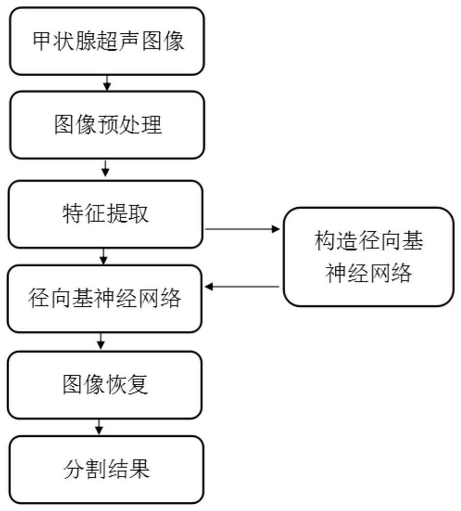
[0082] as Figure 1 As shown, the present embodiment of a radial base neural network based thyroid ultrasound image automatic segmentation method, comprising the following steps:
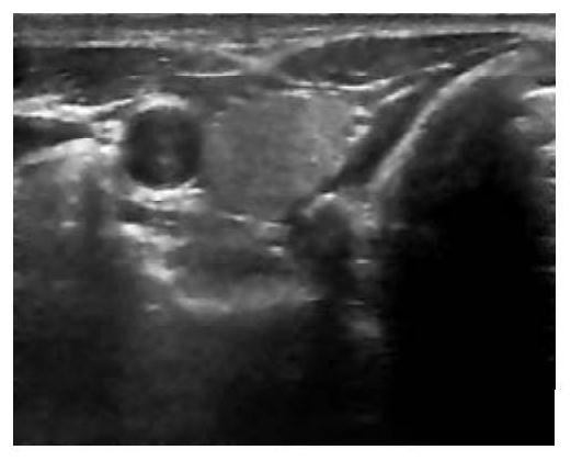
[0083] (1) Enter the original image, as shown in Figure 2(a);
[0084] (2) Pre-processing of the original image;
[0085] (2-1) The original image is noise-reduced using adaptive weight median filtering (AWMF), and the results of the filtering are as shown in Figure 2(b);
[0086] AWMF calculates the weight w for each pixel in the area by counting the local area i,j 。 For the region template of M×M, the point weight coefficient w at the position (i,j). i,j It can be defined as follows:
[0087]
[0088] μ of them x,y and The average of the pixel grayscale values and the variance within the template area, respectively, are the position of the center point of the template area (x, y). g is the scale factor, w 0 is the weight of the template center point (x, y), D is the Euclidean distance from each pixel in the template area to the center point, [·] Represents a rounded function. If the weight is calculated w i,j if it is negative, then this weight is w i,j Set to 0. When calculating the weight value for each pixel in the template area, w i,j After that, the pixel values are sorted from small to large by grayscale intensity, and an ordered sequence can be obtained after the sorting is completed. Pixel value I in this sequence i,jWill be continuous w i,j Second, find the median of this sequence as the grayscale value of the center point pixel. In this example, the 9×9 region template is used, and the parameter is set to w 0 =10 and g=0.25;
[0089] (2-2) Use morphological opening and closing operations to remove the noise generated by using AWMF filters, and the results are as shown in Figure 2(c);
[0090] (2-3) Normalize the input image using histogram equalization, and the result after histogram equalization is as shown in Figure 2(b);
[0091] (3) Features of 16×16 regions in ultrasound images were extracted, and a total of 10 features included local change coefficient (CV), histogram mean (HM), histogram variance (HV), inverse probability block difference (BDIP), normalized scale intensity difference (NMSID), uniformity (GLCMU), inverse moment (GLCMOIDM), contrast ratio (GLCMCON), moment of inertia (GLCMIM), correlation (GLCME)
[0092] (4) The training stage uses the images in the training data set to build a radial base neural network, and the constructed neural network is used for classification during the testing stage; the specific training stage and testing stage are as follows:
[0093] (4-1) During the training phase, the training set contains 850 images of thyroid tissue regions and 1400 non-thyroid tissue regions, each with an image size of 16×16 pixels. Features are extracted from these area images to form a labeled dataset. In the input layer of a radial base neural network, the data is normalized to an input vector with a vector length of 10. The hidden layer is set with 128 nodes, using the Gaussian radial basis function as the basis function of the node, and solving the data center c of each node basis function by determinant i to extend the constant ε i and the weight of the node w i 。 The threshold used for discriminant in the output layer is set to T=0.9.
[0094] (4-2) In the testing stage, on the pre-processed thyroid ultrasound image, the current area is judged to be thyroid or non-thyroid region by sliding window with a size of 16×16 pixels. A pixel that is classified as a thyroid region once or more is labeled as a thyroid region. Slides the window from left to right, top-down, sliding two pixels at a time. After sliding the full frame image, draw the thyroid region according to the table markers, as shown in Figure 3(a) as the input image and Figure 3(b) as the image segmented by the radial base neural network.
[0095](5) Eliminate the area of misclassification and eliminate the aliasing of the split edge; the specific steps are as follows:
[0096] (5-1) Identify the largest area of all the connective areas classified as thyroid glands and reclassify the other areas into non-thyroid tissue regions, as shown in Figure 3(c).
[0097] (5-2) Eliminate the aliasing of the thyroid division boundary, using 4 templates, such as Figure 4 as shown. "×" means that the pixel is not considered, white indicates that the pixel is marked as non-thyroid, and black indicates that the pixel is marked as thyroid. Four templates are used to match the image produced by steps (5-1), and if any of them are matched, the pixel represented by the middle white square is divided into thyroid glands, and the result is shown in Figure 3(d), which is the result of the final segmentation.
[0098] The size of the thyroid ultrasound image used in the present invention is 500×400, a total of 2250 image areas with a size size of 16×16 are used as training samples, comprising 1400 non-thyroid tissue region samples and 850 thyroid tissue region samples. When taking the training samples, the thyroid region is manually segmented and the sample area is extracted using the manual segmentation as the reference standard. During the testing phase, a total of 70 thyroid ultrasound images were used to obtain segmented thyroid ultrasound images and corresponding quantitative indicators. The quantitative indicators are as follows:
[0099]
[0100]
[0101]
[0102]
[0103]
[0104] where TP, TN, FP, FN, A P ,and A N They represent True Positive, Truenegative, False Positive, False Negative, All Positive, and All Negative, respectively. The average quantitative indicators obtained after segmenting 70 images are shown in Table 1.
[0105] Table 1 Test set split quantization results
[0106]
[0107] Figure 5 To comprise the input image, manually segment the image and use the method of the present invention to produce a segmentation result image. wherein (a)(b)(c) is the input image, (d)(e)(f) is an artificially segmented image, (g)(h)(i) is a segmentation result image produced using the method of the present invention.
[0108] It can be seen from the above table that the present invention has a good segmentation effect and accuracy in the segmentation of the ultrasound thyroid region, while simplifying manual operation and reducing manual intervention. The present invention takes the automatic segmentation of the thyroid region as the research object, takes the thyroid ultrasound image as the research object, and focuses on how to automatically segment the thyroid region using a radial base neural network. It is expected to further increase the degree of automation of thyroid computer-aided systems, while automating segmentation, reducing manual workload and intervention, and improving the accuracy of thyroid ultrasound image segmentation.

PUM

no PUM

Description & Claims & Application Information

We can also present the details of the Description, Claims and Application information to help users get a comprehensive understanding of the technical details of the patent, such as background art, summary of invention, brief description of drawings, description of embodiments, and other original content. On the other hand, users can also determine the specific scope of protection of the technology through the list of claims; as well as understand the changes in the life cycle of the technology with the presentation of the patent timeline. Login to view more.

Similar technology patents

Diabetes syndrome prediction system

Owner:CHENGDU UNIV OF TRADITIONAL CHINESE MEDICINE

Classification and recommendation of technical efficacy words

  • Improve identification
  • Good classification effect

Tape cartridge format identification in a single reel tape handling device

InactiveUS6918554B2Improve identificationReduce damagePicture changing apparatusAccessories for auxillary signalsEngineeringMagnetic tape
Owner:QUANTUM CORP
Why Eureka
Try Eureka