ECG waveform simulation method

A simulation method and electrocardiographic wave technology, applied in the field of medical teaching, can solve the problems of limited number of effective electrocardiographic cases for medical students, unfavorable, physical and mental impact of patients, etc., and achieve the effect of enriching clinical teaching cases

Active Publication Date: 2018-09-28
TIANJIN TELLYES SCI
View PDF8 Cites 1 Cited by
  • Summary
  • Abstract
  • Description
  • Claims
  • Application Information

AI Technical Summary

Problems solved by technology

[0009] At present, in the field of medical teaching, medical students follow the teacher to study in the ward or outpatient clinic, and the dynamic electrocardiograph requires the subject to wear the recorder for a long time to record and store

Method used


Image

  • ECG waveform simulation method
  • ECG waveform simulation method
  • ECG waveform simulation method

Examples


example 1

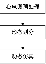
[0092] Instance 1, such as Figure 4 As shown in the ECG simulation method, the specific steps include:

[0093] S1, ECG preprocessing,

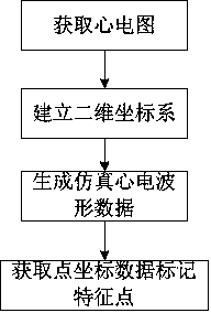
[0094] S11, obtain the clinical electrocardiogram, as shown in A1, it is a normal electrocardiogram waveform,

[0095] S12, establishing a two-dimensional coordinate system,

[0096] S13, generating simulated ECG waveform data, wherein taking the waveform of lead II in Figure A1 as an example, intercepting the waveform of lead II in Figure A1, generating the simulated ECG waveform as shown in A2,

[0097] S14, by linear interpolation method, obtain point coordinate data evenly distributed in time on Figure A2, and mark characteristic points, including P wave start point, QRS wave group start point, T wave end point.

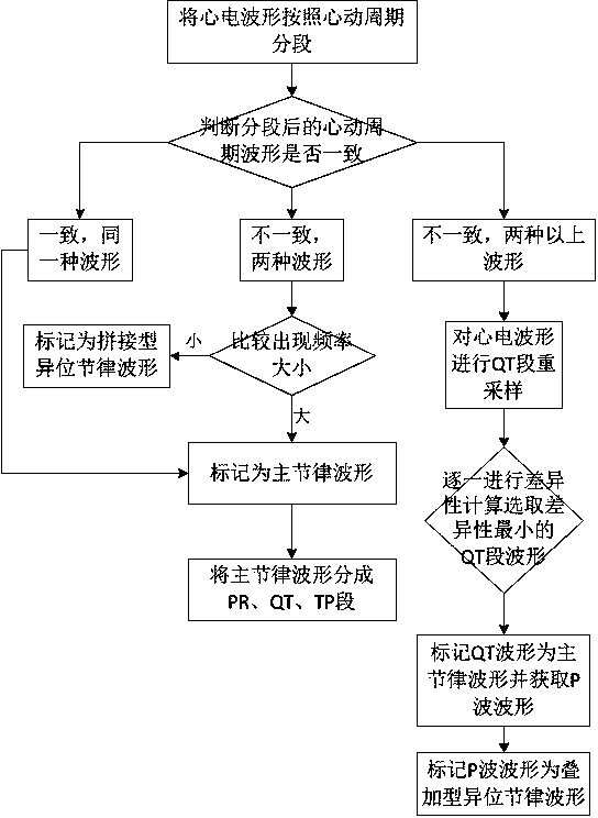
[0098] S2, performing morphological division on the preprocessed ECG according to the waveform features,

[0099] S21, according to the cardiac cycle, the simulated ECG waveform in Figure A2 is segmented, that is, from the ...

example 2

[0112] Instance 2, such as Figure 5 As shown in the ECG simulation method, the specific steps include:

[0113] S1, ECG preprocessing,

[0114] S11, obtain the clinical electrocardiogram, as shown in B1, it is the electrocardiogram waveform of the case of single ventricular premature systole,

[0115] S12, establishing a two-dimensional coordinate system,

[0116] S13, generating simulated ECG waveform data, wherein taking the waveform of lead II in Fig. B1 as an example, intercepting the waveform of lead II in Fig. B1, generating the simulated ECG waveform as shown in B2,

[0117] S14, by linear interpolation method, obtain point coordinate data evenly distributed in time on Figure B2, and mark characteristic points, including P wave start point, QRS wave group start point, T wave end point.

[0118] S2, performing morphological division on the preprocessed ECG according to the waveform features,

[0119] S21, segmenting the simulated ECG waveform in Figure B2 according ...

example 3

[0131] Example 3, such as Image 6 As shown in the ECG simulation method, the specific steps include:

[0132] S1, ECG preprocessing,

[0133] S11, obtain the clinical electrocardiogram, as shown in C1, it is the electrocardiogram waveform of a case of third-degree atrioventricular block and high block,

[0134] S12, establishing a two-dimensional coordinate system,

[0135] S13, generating simulated ECG waveform data, wherein taking the waveform of lead II in Figure C1 as an example, intercepting the waveform of lead II in Figure C1, generating the simulated ECG waveform as shown in C2,


PUM


Abstract

The invention discloses an ECG waveform simulation method, and relates to the field of medical education. The method comprises the steps: 1, acquiring ECG waveform data: acquiring a clinical electrocardiogram, identifying an electrocardiogram curve, obtaining point coordinate data uniformly distributed in the time dimension, and marking a P wave start point, a QRS wave group start point, and a T wave end point; 2, judging the shape of an ECG waveform: segmenting the ECG waveform data according to a cardiac cycle, judging whether all segments of the cardiac cycle waveform after segmentation areconsistent not, marking a main rhythm waveform and an ectopic rhythm waveform, further segmenting the main rhythm waveform, and performing breakpoint smoothing of segmented waveforms; 3, performing dynamic simulation: obtaining a current heart rate by fine-tuning control, calculating the length of the TP segment under an original heart rate and the current heart rate, generating a main rhythm waveform and an ectopic rhythm waveform under the current heart rate, and performing the outputting. According to the present invention, the method achieves the dynamic display of a clinical real electrocardiogram and enriches the clinical teaching examples.

Description

technical field [0001] The invention relates to the field of medical teaching, in particular to a method for simulating an electrocardiogram waveform. Background technique [0002] The heart is the power plant of the human blood circulation. It is precisely because the heart automatically and continuously performs rhythmic contraction and relaxation activities that blood flows continuously in the closed circulatory system and life is maintained. Before and after the heart beats, the heart muscle is excited. During the exciting process, a weak biological current will be generated. In this way, every cardiac cycle of the heart is accompanied by bioelectrical changes. This bioelectrical change can be transmitted to various parts of the body surface. Due to the different tissues of different parts of the body and the different distances from the heart, the electrocardiographic signal exhibits different potentials in different parts of the body. For a normal heart, the direc...

Claims


Application Information

Patent Timeline
IPC IPC(8): G09B23/28
CPCG09B23/28
Inventor 关红彦马培培尚双双张华李南楠
Owner TIANJIN TELLYES SCI
Why Eureka
Try Eureka