
System and method for automated detection and monitoring of dysplasia and administration of chemoprevention

A dysplasia, chemoprevention technology, applied in the preparation of test samples, measuring devices, scientific instruments, etc., can solve the problem of lack of reliable optical tomography to identify precancerous conditions

Inactive Publication Date: 2018-09-28
VISIONGATE
View PDF13 Cites 1 Cited by
  • Summary
  • Abstract
  • Description
  • Claims
  • Application Information

AI Technical Summary

Problems solved by technology

[0010] However, until the disclosure of this paper, there were no reliable methods to use optical tomography to identify precancerous conditions such as dysplasia

Method used


Image

  • System and method for automated detection and monitoring of dysplasia and administration of chemoprevention
  • System and method for automated detection and monitoring of dysplasia and administration of chemoprevention
  • System and method for automated detection and monitoring of dysplasia and administration of chemoprevention

Examples


Embodiment Construction

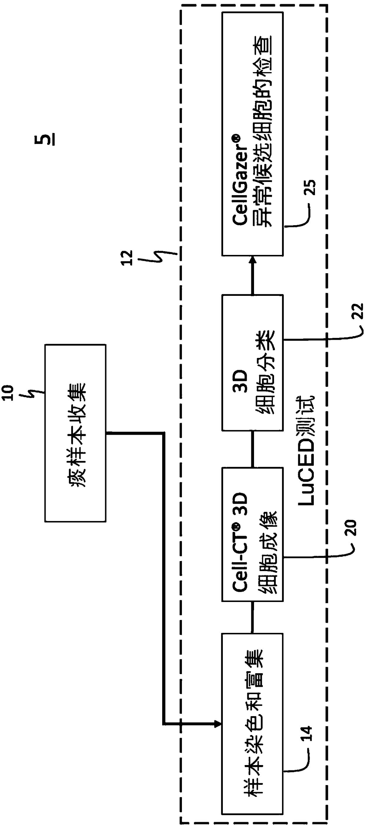
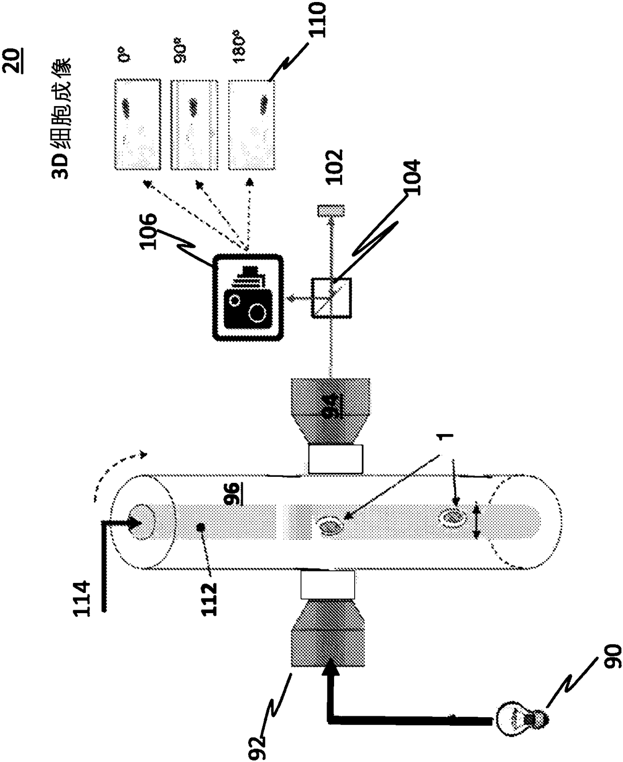
[0025] The following disclosure describes methods for automatic detection and monitoring of dysplasia by analyzing 3D images of cells obtained from sputum samples. Several features of methods and systems according to exemplary embodiments are presented and described in the accompanying drawings. It is to be understood that methods and systems according to other exemplary embodiments may include additional procedures or features different from those shown in the figures. Exemplary embodiments are described herein with respect to an optical tomography unit imaging system. It should be understood, however, that these examples are for illustrative purposes of principles and the invention is not limited thereto.

[0026] The present invention provides an early lung dysplasia and cancer detection system using samples including sputum from a patient processed by an optical tomography system that produces equidistant sub-micron resolution 3D cellular images , and then processed by a...


PUM


Abstract

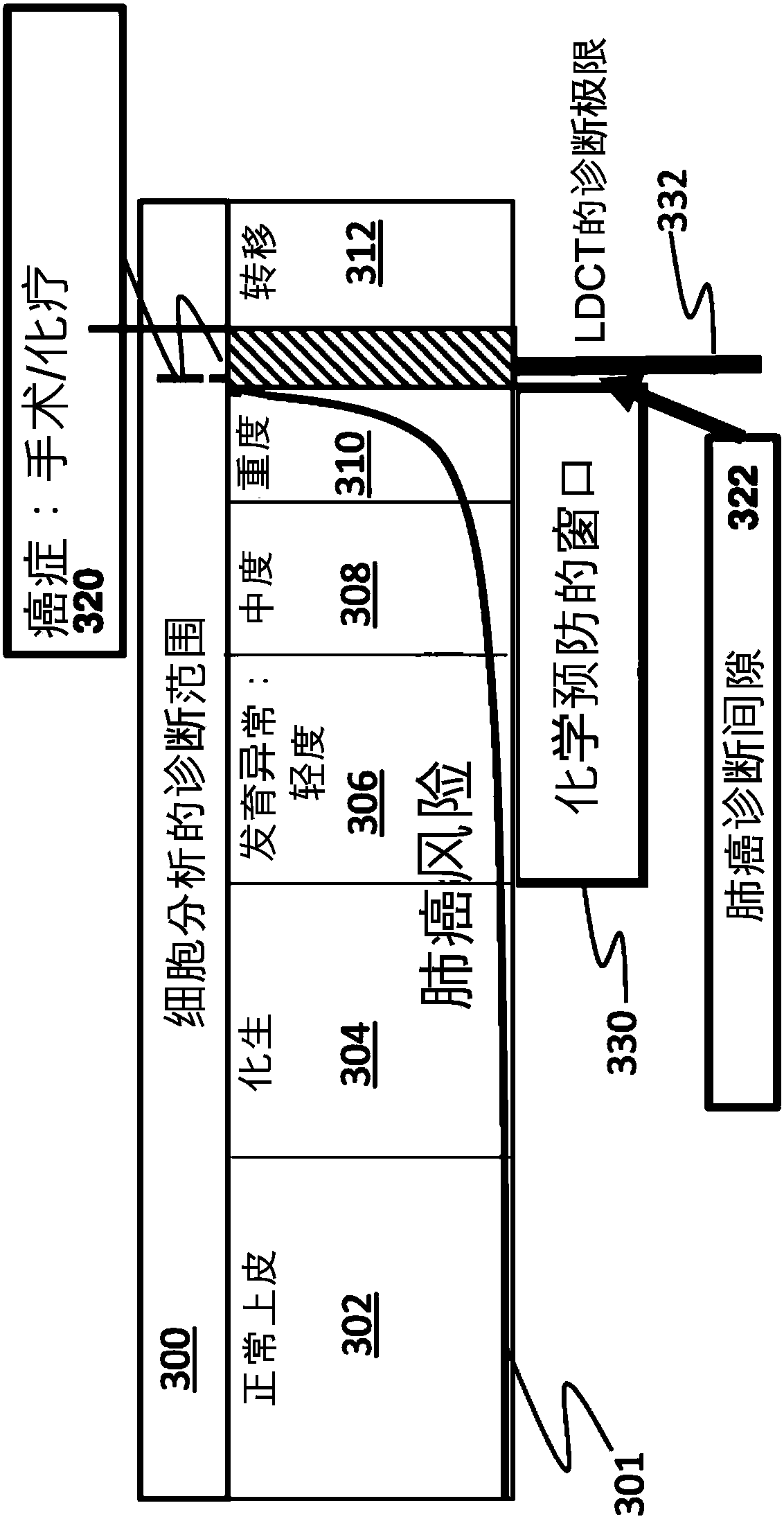
A method for automated detection, monitoring and treatment of dysplasia by analyzing 3D reconstructed images of cells obtained from a specimen includes a biological specimen classifier that classifiescells from the sputum specimen as normal or abnormal. If abnormal cells are detected, then the abnormal cells are further classified as pre-cancerous or cancerous. Pre-cancerous cells are further sub-classified as being of glandular origin or squamous origin (dysplastic cells). This information would be used to define patient therapy so that if the cells are classified as dysplastic, then a cancer chemoprevention pharmaceutical like iloprost is administered to the subject over a predetermined time period to achieve a therapeutic dosage, and if only malignant cells were found or malignant andpre-cancerous cells were found, then next steps would involve patient triage to biopsy and surgery and possibly use of a cancer chemoprevention pharmaceutical.

Description

technical field [0001] The present invention relates to optical tomography at the cellular and subcellular scales. More specifically, the present invention relates to a system and method for determining dysplasia in a sample analyzed by an optical tomography system adapted to detect dysplastic cells, and thereby administering cancer chemopreventive drugs to a patient to reduce developmental abnormalities and reduce lung cancer risk. Background technique [0002] Lung cancer is the second most prevalent cancer in the United States and the deadliest cancer. More than 31 million patients in the United States (US) are at high risk of developing lung cancer, mainly due to age, smoking history, pollution, and other factors, including radon exposure, family history of lung cancer, and others. Approximately 160,000 American patients die from lung cancer each year. As of this writing, lung cancer can only be cured with surgery if it is detected at an early stage (mainly stage I an...

Claims


Application Information

Patent Timeline
Patent Type & Authority Applications(China)
IPC IPC(8): G01N33/48G01N33/483G01N33/487G01N21/64A61P35/00A61P35/04A61B8/13
CPCG01N1/40G01N2015/1445G01N33/5011G01N33/5026G01N2800/7028A61P35/00A61P35/04G01N15/1433G01N15/149
Inventor 艾伦·C·纳尔逊迈克尔·G·迈耶
Owner VISIONGATE
Learn More