Method for judging rupture risk of reconstructed ear scaffold

A technology for reconstructing ears and ulceration, which is applied in image data processing, medical science, 3D modeling, etc., and can solve problems such as skin ulceration and stent exposure

Active Publication Date: 2018-11-02
SOUTHERN MEDICAL UNIVERSITY
View PDF8 Cites 1 Cited by
  • Summary
  • Abstract
  • Description
  • Claims
  • Application Information

AI Technical Summary

Problems solved by technology

Clinicians have conducted a lot of research on how to sculpt a realistic outer auricle, but they have ignored...

Method used


Image

  • Method for judging rupture risk of reconstructed ear scaffold
  • Method for judging rupture risk of reconstructed ear scaffold
  • Method for judging rupture risk of reconstructed ear scaffold

Examples


Embodiment 1

[0058] A method for judging the rupture risk of a reconstructed ear support, comprising the steps of:

[0059] S1, scanning the initial body of the ear stent to obtain contour data of the initial body of the ear stent;

[0060] S2, importing the solid model of the STL format of the ear support initial body obtained in step S1 into the geomagicFreeFrom&touch X software for surface repair, and saving the solid model after the surface repair as STL format;

[0061] S3, import the STL format saved in the solid model after the surface restoration in step S2 into Geomagic Wrap software, and the selection commands are: precise surface—automatic surfacing—constructing contour lines—constructing surface patches—constructing grids—fitting surfaces, and finally Generate Nurbs surface, export the generated Nurbs surface and save it in IGES format;

[0062] S4, import the Nurbs surface in IGES format obtained in step S3 into the HyperMesh14.0 software to establish a finite element model, ...

Embodiment 2

[0090] The method of the present invention will be further described with the application of a clinically sculpted ear holder model.

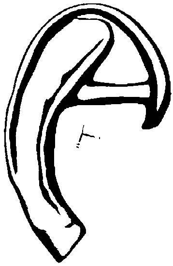
[0091] The ear support model of this embodiment is aimed at a 7-year-old male child with congenital right microtia incomplete, the incomplete ear is in the shape of a hillock, and the classification type of ear deformity is third degree. For the repair of its outer ear, ear brackets have been carved, such as figure 1 shown, but the risk of collapse is unknown. The method of the present invention is used to judge the risk of rupture of the ear support.

[0092] The specific process is as follows:

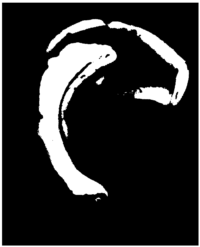
[0093] S1, will figure 1 Put the carved ear bracket on the sterile table, and paste the sterilized positioning target points around it. Use the handscan700 handheld 3D scanner to scan with an accuracy of 0.03mm and a measurement rate of 480,000 times per second to obtain the ear bracket Contour data, such as figure 2 shown;

[0094] S2, import ...

Embodiment 3

[0120] The ear support model of this embodiment is for a 7-year-old male child with congenital right microtia in Example 2, the residual ear is hillock-shaped, and the classification type of ear deformity is third degree. The Medpor model was selected specifically for its external ear repair. The Medpor model was purchased from Strker’s right ear implants represented by Chengdu Qintian Biotechnology Co., Ltd., the models are 8328 and 8330; 64 rows of 128-slice CT scans were used to confirm that the internal structure of the ear implant is basically solid, and the external structure uses RexcanDS3 blue light A fully automatic 3D scanner scans it to obtain the original data. The method of the present invention is used to analyze the risk of rupture of the ear support to determine whether optimization and improvement are needed.

[0121] The specific process is as follows:

[0122] S, conduct a risk analysis of ear stent rupture, optimize and adjust the ear stent according to t...


PUM


Abstract

The invention discloses a method for judging the rupture risk of a reconstructed ear scaffold. The method comprises the following steps that: S1: scanning the ear scaffold, and obtaining the outline data of the ear scaffold; S2: importing an entity model into geomagic FreeFrom&touch X software to carry out hook face repairing; S3: generating a Nurbs hook surface; S4: importing the IGES (The Initial Graphics Exchange Specification) format of S3 into HyperMesh14.0 software to establish a finite element model; S5: importing a three-dimensional finite element ear model which is obtained in S4 andcontains cartilages and different skin thicknesses into finite element specialty analysis software Abaqus to carry out finite element analysis; and S6: obtaining a stress-strain analysis result of cartilages, skin, cartilage deformation and skin deformation, and judging the rupture risk of the ear scaffold according to the finite element stress result. By use of the method, a situation that the rupture may happen when the ear scaffold is used can be evaluated in advance, and a rupture possibility is lowered.

Description

technical field [0001] The invention relates to the technical field of 3D plastic digital medicine, in particular to a method for judging the risk of rupture of a reconstructed ear support. Background technique [0002] The incidence of microtia is increasing significantly all over the world. It was recently reported that the incidence in my country is 5.18 / 10000, which is a substantial increase compared with the past. There are more men than women, most of which are unilateral, and the incidence rate on the right side is higher than that on the left side. The outer auricle is located on both sides of the skull, and is often overlooked because it does not have specific functions. However, due to the lack of ears of patients, in addition to being unable to wear glasses, masks and other accessories, they are often ridiculed in appearance, causing low self-esteem. Part of the external auditory canal is blocked. It may affect the patient's hearing, increase the physical and psyc...

Claims


Application Information

Patent Timeline
IPC IPC(8): G06T17/20A61F2/50
CPCA61F2/50A61F2002/5001G06T17/205G06T2207/30052
Inventor 黄文华钟静
Owner SOUTHERN MEDICAL UNIVERSITY
Why Eureka
Try Eureka