
Self-portrait shared fundus camera device

A camera device and Selfie technology, applied in the field of shared medical care, can solve problems such as failure to convert image transmission formats, no self-portrait fundus camera, and fundus image data that cannot be shared and viewed, so as to save medical resources and patient costs, and benefit Rational use and the effect of exempting medical personnel from operating

Active Publication Date: 2020-07-10
NANTONG UNIVERSITY
View PDF4 Cites 0 Cited by
  • Summary
  • Abstract
  • Description
  • Claims
  • Application Information

AI Technical Summary

Problems solved by technology

On the basis of the traditional fundus camera, functions such as non-mydriasis, automatic focus, automatic shooting, automatic image optimization, and automatic printing have been developed. The adjustable flash intensity greatly improves the comfort and cooperation of patients [Chen Jian , Zheng Shaohua, Pan Lin, Yu Lun. Coarse-fine autofocus method in non-mydriatic fundus cameras [J]. Acta Optics Sinica, 2014, 34(11): 117-128.】It can also realize remote transmission of images, but The image collection is basically machine-assisted auto-focusing supplemented by manual shooting by the operator. The fundus image data in different hospitals has not been shared and viewed, there is no Selfie fundus camera, and the image transmission format has not been converted to DICOM medical images. Standard format, making medical imaging data better shared in medical institutions

Method used


Image

  • Self-portrait shared fundus camera device
  • Self-portrait shared fundus camera device
  • Self-portrait shared fundus camera device

Examples


Embodiment Construction

[0031] In order to make the purpose, technical solutions and advantages of the embodiments of the present invention clearer, the technical solutions in the embodiments of the present invention will be clearly and completely described below in conjunction with the drawings in the embodiments of the present invention. Obviously, the described embodiments It is a part of embodiments of the present invention, but not all embodiments. Based on the embodiments of the present invention, all other embodiments obtained by persons of ordinary skill in the art without making creative efforts belong to the protection scope of the present invention.

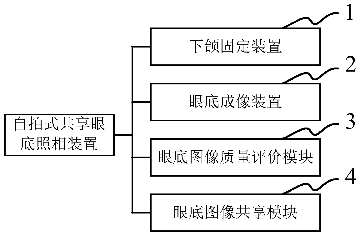
[0032] Such as figure 1 As shown, this embodiment provides a self-portrait shared fundus photography device, which includes: a jaw fixation device 1 , a fundus imaging device 2 , a fundus image quality evaluation module 3 and a fundus image sharing module 4 .

[0033] The mandibular fixation device 1 is used to fix the mandible of the self-e...


PUM


Abstract

The invention relates to a self-timer type shared fundus photography device. The self-timer type shared fundus photography device comprises a lower jaw fixing device for fixing the lower jaw of a self-examiner, a fundus imaging device for taking a fundus image of the self-examiner by adopting a visual target focusing and automatic focusing technology and receiving personal information input of theself-examiner, a fundus image quality evaluation module for judging whether the fundus image meets preset requirements, displaying the fundus image for the self-examiner to choose to submit or retakewhen the fundus image meets the preset requirements, and converting the fundus image into a fundus image with a set file format when a submission instruction is selected, and a fundus image sharing module for storing the personal information of the self-examiner and the corresponding fundus image converted into the set file format for sharing and calling. The self-timer type shared fundus photography device provided by the invention can realize self-photographing of fundus images of patients, avoids operations of medical personnel, realizes digital shared storage and retrieval of fundus imagedata and report information between hospitals, greatly reduces repeated examination, saves medical resources and patient expenses, and is beneficial to rational utilization of medical information.

Description

technical field [0001] The invention relates to the field of shared medical technology, in particular to a self-portrait sharing fundus camera device based on IHE-XDS integration technology. Background technique [0002] The fundus is the tissue at the inner back of the eyeball, including: the retina, optic disc, macula, and retinal blood vessels. Patients suffering from hypertension, diabetes, kidney disease and other diseases can be detected and diagnosed early through fundus examination. Fundus imaging examination is the most basic and important inspection method in the field of ophthalmology, and fundus camera is an important means to record retinopathy. Carrying out fundus camera general inspection is of great significance for early detection and early diagnosis of hypertension, diabetes and other ophthalmic diseases. At present, my country's medical resources are relatively insufficient, especially in remote rural areas. The shared fundus examination and medical image...

Claims


Application Information

Patent Timeline
Patent Type & Authority Patents(China)
IPC IPC(8): A61B3/12A61B3/14
CPCA61B3/12A61B3/14
Inventor 吴辉群于镆铘张晓峰董建成耿兴云
Owner NANTONG UNIVERSITY
Learn More