Method for recognizing spatial difference by utilizing complex number fMRI (functional magnetic resonance imaging) space source phase

A phase recognition and spatial difference technology, applied in the field of biomedical signal processing, to achieve high sensitivity and reliability

Active Publication Date: 2018-11-30
DALIAN UNIV OF TECH
View PDF6 Cites 10 Cited by
  • Summary
  • Abstract
  • Description
  • Claims
  • Application Information

AI Technical Summary

Problems solved by technology

However, does the spatial source phase also convey unique information about internal

Method used


Image

  • Method for recognizing spatial difference by utilizing complex number fMRI (functional magnetic resonance imaging) space source phase
  • Method for recognizing spatial difference by utilizing complex number fMRI (functional magnetic resonance imaging) space source phase
  • Method for recognizing spatial difference by utilizing complex number fMRI (functional magnetic resonance imaging) space source phase

Examples


Embodiment Construction

[0033] A specific embodiment of the present invention will be described in detail below in conjunction with the technical scheme and accompanying drawings.

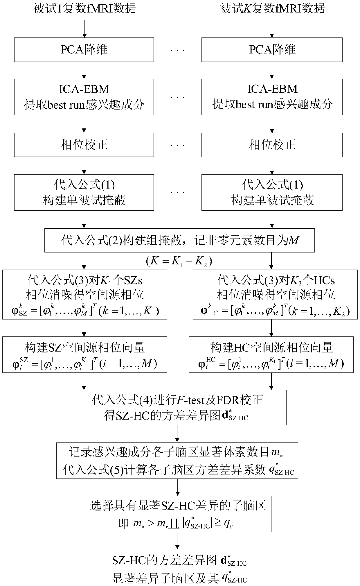
[0034] Existing K 1 = 42 SZs and K 2= Complex fMRI data acquired in the resting state of 40 HCs (K=82). In the time dimension, T=146 scans were performed, each scan obtained 53×63×46 whole brain data, and the number of voxels in the brain was V=62336. The steps of using the present invention to analyze the spatial source phase to identify the spatial difference between SZs and HCs are as attached figure 2 shown.

[0035] Step 1: Input multi-subject complex fMRI data

[0036] Step 2: For all single subjects X k Carry out PCA dimensionality reduction, take N=120, and obtain dimensionality reduction data

[0037] Step 3: Use the complex EBM algorithm for all single subjects Perform R=10 times of complex ICA separation in sequence, extract the component of interest DMN from 120 estimated components, use the best ...


PUM


Abstract

The invention relates to a method for recognizing a spatial difference by utilizing a complex number fMRI (functional magnetic resonance imaging) space source phase, belonging to the technical field of biomedical signal processing. The method comprises the following steps of carrying out spatial ICA (independent component analysis) separation on complex number fMRI data, choosing an interesting ingredient, then performing phase correction and noise elimination to obtain the spatial source phase of the interesting ingredient, and finally, calculating through a variance homogeneity analysis anda variance difference coefficient so as to recognize the differences of SZs and HCs in the spatial source phase. Compared with widely applied amplitude data, the method utilizes the spatial source phase to recognize the spatial difference between a schizophrenia patient group and a healthy control group, and can display higher flexibility and reliability. For example, specific to complex number fMRI data of 82 testees in a quiescent condition, the spatial source phase is used for extracting differential voxel, which is increased by more than 122.95% in DMN, on the premise of not using FDR correction, and the differential voxel, which is extracted from the spatial source phase and can pass the FDR correction, is 69% more than the amplitude.

Description

technical field [0001] The present invention relates to the field of biomedical signal processing, in particular to a method for identifying schizophrenia patients (patients with schizophrenia, SZs) and healthy controls using the spatial source phase of complex functional magnetic resonance imaging (fMRI) data A method for spatially activated brain region (spatial map (SM)) differences between groups (healthy controls (HCs)). Background technique [0002] fMRI is a neurological imaging modality based on blood-oxygenation-level-dependent effects, which can reflect the internal activities of the brain. Due to its non-invasive and high spatial resolution, fMRI technology has been widely used in many fields, such as basic neuroscience research and clinical medicine, etc., and is one of the important tools for understanding human brain function and studying how diseases disrupt brain activity . Independent component analysis (ICA) is a typical data-driven blind source separatio...

Claims


Application Information

Patent Timeline
IPC IPC(8): A61B5/055
CPCA61B5/0037A61B5/0042A61B5/055A61B5/7203A61B5/748A61B2576/026
Inventor 林秋华邱悦
Owner DALIAN UNIV OF TECH
Why Eureka
Try Eureka