MIL-based medical image lesion point accurate marking method

A medical image and lesion technology, applied in the field of medical image processing, can solve the problems of scarce samples, precise marking of lesion areas, and small number, and achieve the effects of reducing workload, high universality, and high efficiency

Inactive Publication Date: 2018-11-30
SOUTHWEST JIAOTONG UNIV
View PDF4 Cites 9 Cited by
  • Summary
  • Abstract
  • Description
  • Claims
  • Application Information

AI Technical Summary

Problems solved by technology

[0002] The images in the medical field are marked as: tumors, nodules, calcifications and other lesion areas in image data such as DR films and CT films are marked by doctors in dicom and other image data in the form of human-computer interaction; with deep learning in For the expansion in the field of medical image processing, training requires large-scale standard data sets to support; therefore, the problem of accurate labeling of a large number of data sets is particularly important; different from the labeling of ordinary images, the labeling of medical images requires professional knowledge and skills Therefore, it is difficult to obtain a large amount of accurately labeled data sets for deep neural network learning; at present, the traditional deep learning is widely used in the field of medical images, and there are still the following problems: First, the precise labeling of images in the medical field requires high It is not meaningful for non-medical personnel to label medical images; but objectively, doctors in our country have a high workload and it is difficult to have the energy to accurately label medical images; and there are a large number of different types of lesions in reality images, but many stored images have not been marked and used in time, and the resource utilization rate is low; second, even if doctors can accurately mark medical images, because data labeling needs to eliminate personal subjectivity, it requires multiple people to label. Take the comprehensive average result; this leads to a small amount of effective labeling data that can reach the application level; thirdly, the disease image samples are limited by the incidence of the disease, some diseases are rare because of the rare disease, it is difficult to The problem of constructing a data set of sufficient magnitude
[0003] At present, the reform of digital medical records has been extensively carried out in existing medical institutions, and many PACS systems and other medical record databases containing medical imaging data, biochemical data, etc. have been built; these medical records contain diagnostic conclusions, but do not accurately mark the lesion area; currently Most of the medical image data are inaccurately labeled data (only marked with and without disease), but the location coordinates of the lesion are not given accurately; therefore, it is difficult for the current deep learning algorithm to directly adapt to

Method used


Image

  • MIL-based medical image lesion point accurate marking method
  • MIL-based medical image lesion point accurate marking method

Examples


Embodiment Construction

[0023] The present invention will be further described below in conjunction with the accompanying drawings and specific embodiments.

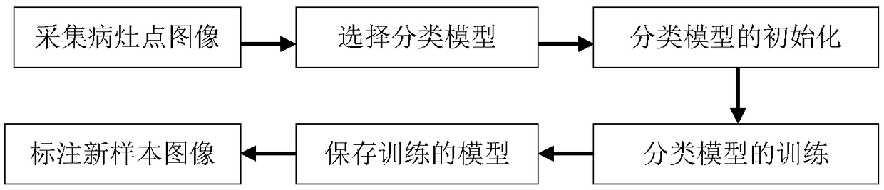
[0024] A method for accurately labeling medical image focus points based on MIL, comprising the following steps:

[0025] Step 1: Collect medical image data sets, which are divided into positive sample sets and negative sample sets;

[0026] Collect several images of different types of lesion points through the camera of medical equipment, and select images with clear pictures at a certain interval of images as the original data set; select a certain number of pictures, and make corresponding image labels for subsequent training.

[0027] Step 2: Initialize the classification model;

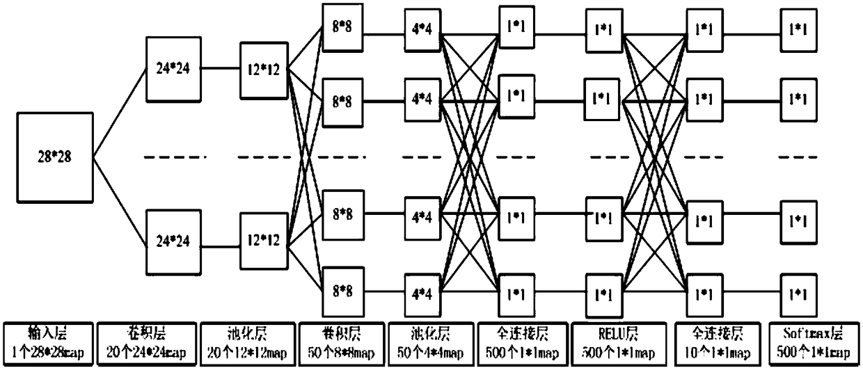
[0028] Select samples with labels for supervised learning, and initialize the classification model; the classification model chooses a model based on the LeNet-based accurate classification of medical image lesions. The classification model is not limited to LeNe...


PUM


Abstract

The invention discloses an MIL-based medical image lesion point accurate marking method. The MIL-based medical image lesion point accurate marking method comprises the following steps: a step 1: acquiring a medical image data set which is divided into a positive sample set and a negative sample set; a step 2: performing initialization of a classification model; a step 3: acquiring S instances foreach sample in the positive sample set, and inputting each instance into the classifier model; recording an instance with the maximal lesion point probability value, and then adding the instance to adata set D; a step 4 : acquiring S instances for each sample in the negative sample set, inputting each instance into the classifier model, recording an instance which is less likely to have a lesionpoint, and adding the instance into the data set D; a step 5: performing iterative training in the neural network classifier model to obtain a training model; and a step 6: detecting and marking new samples according to the classification model. The method can accurately mark the lesion point samples in large quantities, is fast in speed and low in cost, and is extremely efficient.

Description

technical field [0001] The invention relates to a medical image processing method, in particular to an MIL (Multiple Instance Learning)-based method for accurately labeling medical image focus points. Background technique [0002] The images in the medical field are marked as: tumors, nodules, calcifications and other lesion areas in image data such as DR films and CT films are marked by doctors in dicom and other image data in the form of human-computer interaction; with deep learning in For the expansion in the field of medical image processing, training requires large-scale standard data sets to support; therefore, the problem of accurate labeling of a large number of data sets is particularly important; different from the labeling of ordinary images, the labeling of medical images requires professional knowledge and skills Therefore, it is difficult to obtain a large amount of accurately labeled data sets for deep neural network learning; at present, the traditional deep...

Claims


Application Information

Patent Timeline
Patent Type & Authority Applications(China)
IPC IPC(8): G16H30/20G06N3/04
CPCG16H30/20G06N3/045
Inventor 唐鹏万加龙金炜东
Owner SOUTHWEST JIAOTONG UNIV
Why Eureka
Try Eureka