Method, device, device and medium for suppressing cerebrospinal fluid signal in blood vessel wall imaging

A blood vessel wall and cerebrospinal fluid technology, applied in the field of cerebrospinal fluid signal suppression in vascular wall imaging, can solve problems such as the inability to provide cerebrospinal fluid signal suppression methods, low recognition of the outer boundary of the blood vessel wall, and incomplete suppression of cerebrospinal fluid signals, etc., to facilitate diagnosis and Quantitative analysis, improvement of suppression effect, effect of improvement of tissue contrast

Active Publication Date: 2020-05-22
SHANGHAI UNITED IMAGING HEALTHCARE
View PDF12 Cites 0 Cited by
  • Summary
  • Abstract
  • Description
  • Claims
  • Application Information

AI Technical Summary

Problems solved by technology

[0004] The purpose of the present invention is to provide a method, device, equipment and storage medium for suppressing cerebrospinal fluid signals in vessel wall imaging, aiming to solve the problem of magnetic The problem of incomplete suppression of cerebrospinal fluid signal and low recognition of the outer boundary of the vessel wall in resonance vessel wall imaging

Method used


Image

  • Method, device, device and medium for suppressing cerebrospinal fluid signal in blood vessel wall imaging
  • Method, device, device and medium for suppressing cerebrospinal fluid signal in blood vessel wall imaging
  • Method, device, device and medium for suppressing cerebrospinal fluid signal in blood vessel wall imaging

Examples


Embodiment 1

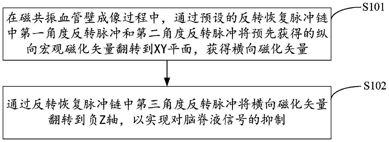
[0021] figure 1 It shows the implementation process of the method for suppressing cerebrospinal fluid signal in the imaging of the vessel wall provided by Embodiment 1 of the present invention. For the convenience of description, only the parts related to the embodiment of the present invention are shown, and the details are as follows:

[0022] In step S101, during the magnetic resonance vessel wall imaging process, the pre-acquired longitudinal macroscopic magnetization vector is reversed to the XY plane through the first angle inversion pulse and the second angle inversion pulse in the preset inversion recovery pulse chain, Obtain the transverse magnetization vector.

[0023] Embodiments of the present invention are applicable to medical image processing platforms, systems or devices, such as personal computers, servers, and the like. In the process of magnetic resonance vessel wall imaging, after the human body enters the main magnetic field of the magnetic resonance syst...

Embodiment 2

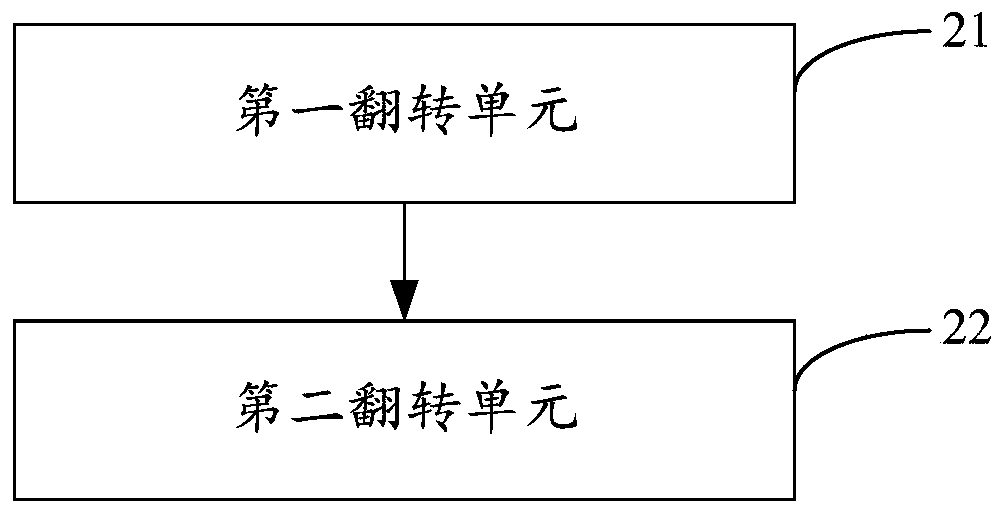
[0032] figure 2 The structure of the cerebrospinal fluid signal suppression device in the blood vessel wall imaging provided by the second embodiment of the present invention is shown. For the convenience of description, only the parts related to the embodiment of the present invention are shown, including:

[0033] The first inversion unit 21 is configured to invert the pre-acquired longitudinal macroscopic magnetization vector to In the XY plane, the transverse magnetization vector is obtained.

[0034]Embodiments of the present invention are applicable to medical image processing platforms, systems or devices, such as personal computers, servers, and the like. In the process of magnetic resonance vessel wall imaging, after the human body enters the main magnetic field of the magnetic resonance system, it is refocused by launching a preset imaging sequence (for example, a fast spin echo imaging sequence or a fast spin echo imaging sequence with a variable flip angle). Pul...

Embodiment 3

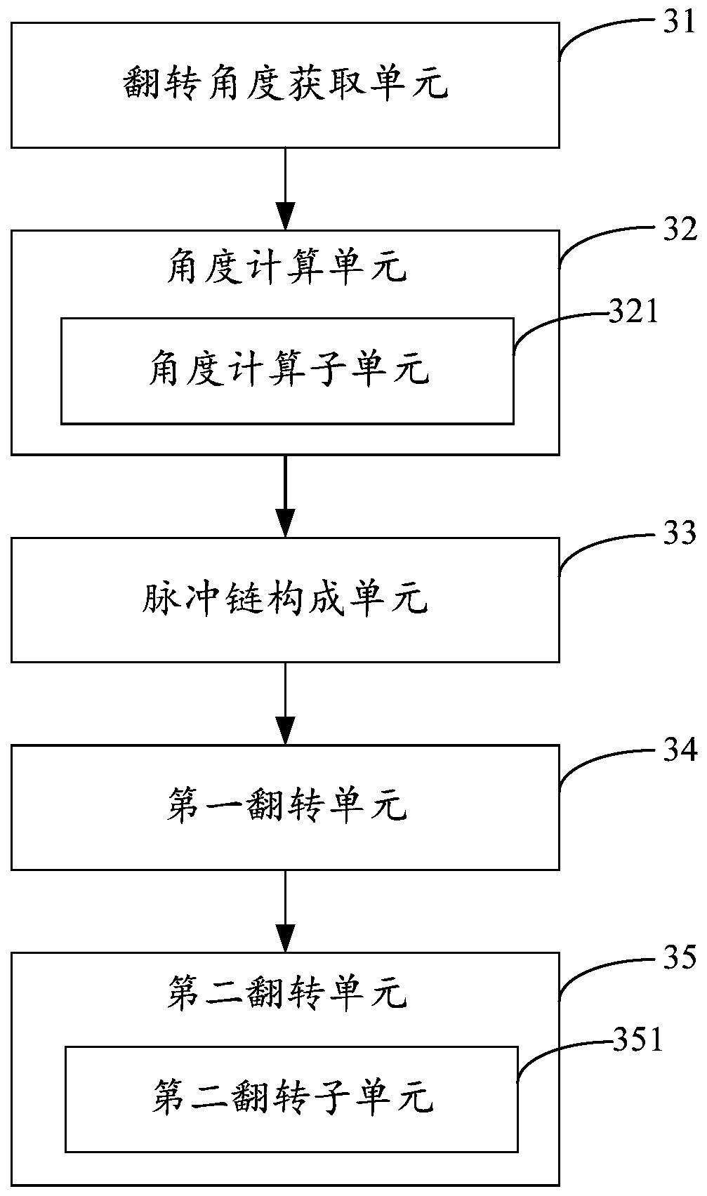
[0040] image 3 The structure of the device for suppressing the cerebrospinal fluid signal in the imaging of the vessel wall provided by the third embodiment of the present invention is shown. For the convenience of description, only the parts related to the embodiment of the present invention are shown, including:

[0041] The flip angle acquiring unit 31 is configured to acquire the flip angle corresponding to the last refocusing pulse in the previously transmitted imaging sequence refocusing pulse chain during the magnetic resonance vessel wall imaging process.

[0042] Embodiments of the present invention are applicable to medical image processing platforms, systems or devices, such as personal computers, servers, and the like. Magnetic resonance vessel wall imaging is to generate echo signals by transmitting fast spin echo imaging sequences or fast spin echo imaging sequences with variable flip angles, and then generate magnetic resonance vessel wall images based on the g...


PUM


Abstract

Disclosed are a method and apparatus for inhibiting a cerebrospinal fluid signal in vascular wall imaging, and a device and a medium. The method comprises: during magnetic resonance vascular wall imaging, flipping a pre-obtained longitudinal macroscopic magnetization vector to an XY plane through a first angle inversion pulse and a second angle inversion pulse in a preset inversion recovery pulse chain, so as to obtain a transverse magnetization vector (S101); and flipping the transverse magnetization vector to a negative Z axis through a third angle inversion pulse in the inversion recovery pulse chain, so as to realize the inhibition of a cerebrospinal fluid signal (S102). The effect of inhibiting a cerebrospinal fluid signal in magnetic resonance vascular wall imaging is improved, and the tissue contrast of a vascular wall image is thus also improved, thereby facilitating diagnosis and quantitative analysis carried out by a doctor.

Description

technical field [0001] The invention belongs to the technical field of magnetic resonance imaging, and in particular relates to a method, device, equipment and medium for suppressing cerebrospinal fluid signals in blood vessel wall imaging. Background technique [0002] Cardiovascular and cerebrovascular diseases have become the primary disease that threatens human health, and atherosclerotic plaque is an important risk factor leading to the occurrence of cardiovascular and cerebrovascular diseases. Stroke events are of great significance. Since intracranial arteries are surrounded by cerebrospinal fluid, in order to clearly distinguish the boundaries of the intracranial artery wall and the plaque attached to it, it is necessary to simultaneously suppress arterial blood flow and cerebrospinal fluid signals. At present, Delays Alternating with Nutation for Tailored Excitation (DANTE) can effectively suppress arterial blood flow and cerebrospinal fluid signals. This technolog...

Claims


Application Information

Patent Timeline
Patent Type & Authority Patents(China)
IPC IPC(8): G01R33/563A61B5/055
CPCA61B5/055G01R33/5635
Inventor 郑海荣刘新张娜张磊贾琳贾文霄邹超梁栋万丽雯赵世华
Owner SHANGHAI UNITED IMAGING HEALTHCARE
Why Eureka
Try Eureka