Cervical cancer histopathology image analysis method and device based on depth learning

A technology of deep learning and image analysis, applied in the medical field, can solve the problems of poor transferability, processing, and inability to diagnose the degree of cancer differentiation of patients globally, so as to achieve the effect of auxiliary diagnosis and enhanced intelligence

Inactive Publication Date: 2019-02-22
四川智动木牛智能科技有限公司
View PDF5 Cites 22 Cited by
  • Summary
  • Abstract
  • Description
  • Claims
  • Application Information

AI Technical Summary

Problems solved by technology

[0009] However, this method can only select a single cell histopathological image with low noise and high definition for processing, and cannot directly process the complete histopathological microsection image, and needs to manually extract the single cell image; this method uses 27 artificially designed morphological features are used as the input value of machine learning. This method has poor transferability and is prone to overfitting in learning; and it can only classify the cell type of a single cell, and cannot diagnose the patient's cancer globally. The degree of differentiation, thus assisting in judging the patient's cancer condition

Method used


Image

  • Cervical cancer histopathology image analysis method and device based on depth learning
  • Cervical cancer histopathology image analysis method and device based on depth learning
  • Cervical cancer histopathology image analysis method and device based on depth learning

Examples


Embodiment Construction

[0044] All features disclosed in this specification, or steps in all methods or processes disclosed, may be combined in any manner, except for mutually exclusive features and / or steps.

[0045]Any feature disclosed in this specification, unless specifically stated, can be replaced by other alternative features that are equivalent or have similar purposes. That is, unless expressly stated otherwise, each feature is one example only of a series of equivalent or similar features.

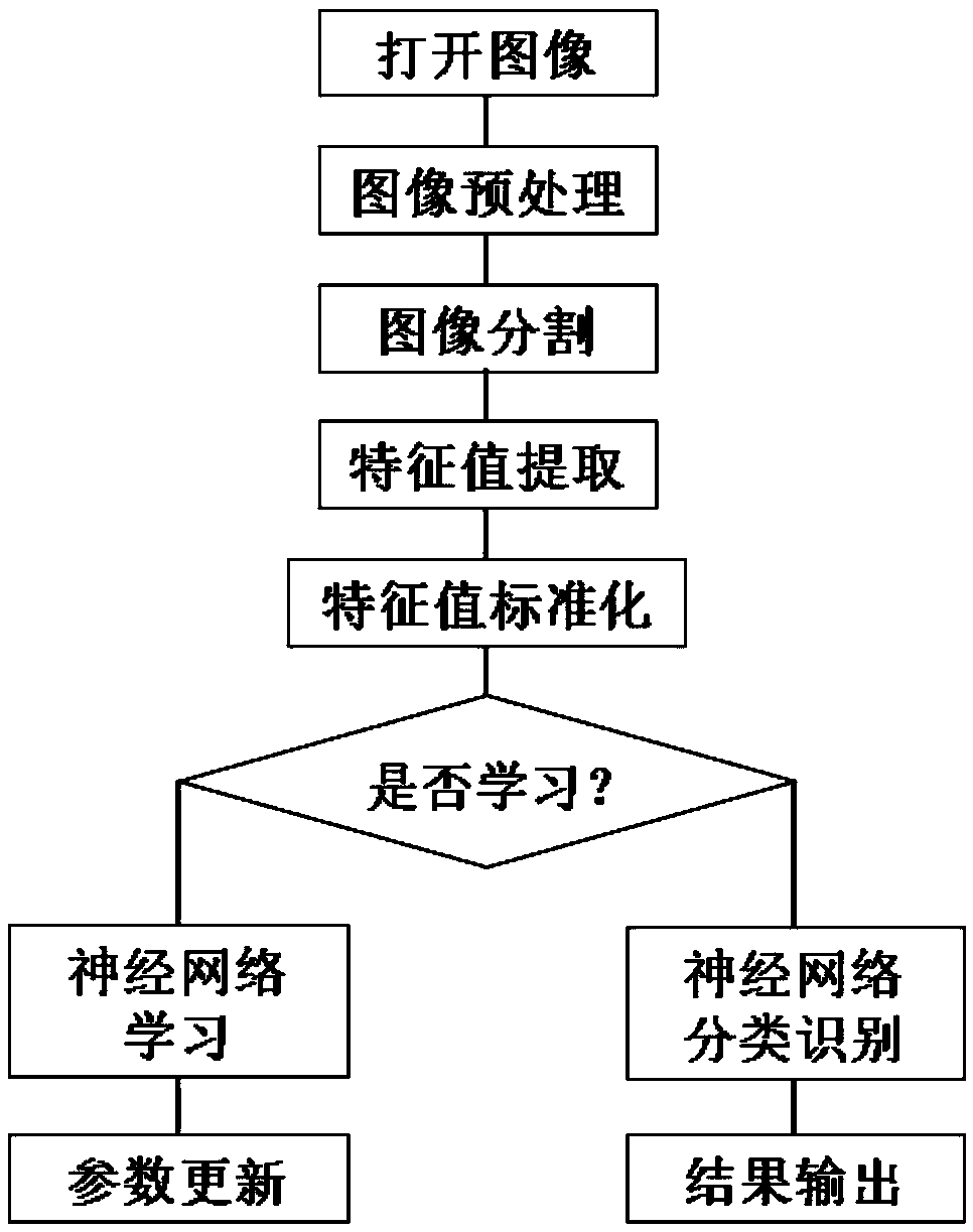
[0046] The specific scheme of the cervical cancer histopathological image analysis method based on deep learning provided by the present invention is as follows:

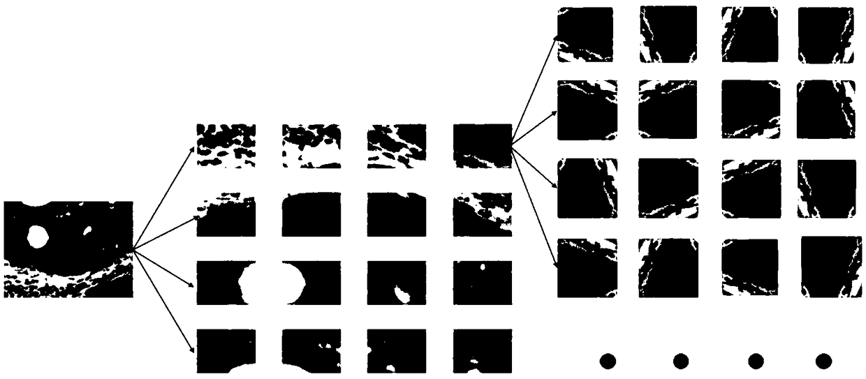
[0047] 1. Sample data acquisition and enhancement

[0048] Histopathological microscopic images of cervical cancer tissue sections were prepared by the Department of Pathology, China Medical University, and the tumor pathological type, degree of differentiation, and tumor size were recorded. Immunohistochemical staining was performed using...


PUM


Abstract

The invention discloses a cervical cancer histopathology image analysis method and device based on depth learning. The method comprises the following steps: acquiring cervical cancer histopathology images and setting image labels for each image; training two convolution neural networks respectively based on the images used for training, and obtaining the two trained convolution neural networks; fixing parameters of the two trained convolutional neural networks, and training the all-connected layer one based on the images used for training, so that the trained all-connected layer one is obtained. The image to be tested is input into the trained classifier, and the two convolution neural networks extract feature vectors from the image respectively. The output feature vectors f1 and f2 are spliced together and input to the full connection layer 1, and the output feature vector f3. The classification result is determined by the element with the largest value in the feature vector f3 The invention can automatically classify and identify the differentiation degree of the original histopathological slice micrograph collected by the doctor and assist the doctor in diagnosis.

Description

technical field [0001] The present invention relates to the medical field, in particular to a method and device for analyzing cervical cancer histopathological images based on deep learning. Background technique [0002] Cervical cancer is one of the common gynecological malignancies. For now, research on computer-aided diagnosis of histopathological microscopic images of cervical cancer mainly focuses on the classification of cervical histopathological images using classical image feature extraction methods and machine learning classification methods. Image segmentation and pathological abnormality screening, few studies have conducted computer-aided diagnosis research on the degree of differentiation of cervical cancer histopathological images. [0003] The prior art is based on using computer vision methods to extract features from histopathological images of only a single cervical cancer cell, and then using traditional machine learning methods to further classify the ex...

Claims


Application Information

Patent Timeline
Patent Type & Authority Applications(China)
IPC IPC(8): G06K9/62G06N3/04G06T7/00G06T7/136
CPCG06T7/0012G06T7/136G06T2207/30096G06N3/045G06F18/241
Inventor 李晨孔繁捷蒋涛许宁
Owner 四川智动木牛智能科技有限公司
Why Eureka
Try Eureka