Antibody capable of binding tigit or antigen-binding fragment thereof and use thereof
An antibody and application technology, applied in the field of antibodies to achieve the effect of inhibiting tumor growth
- Summary
- Abstract
- Description
- Claims
- Application Information
AI Technical Summary
Problems solved by technology
Method used
Image
Examples
Embodiment 1
[0158]Example 1: Preparation of Antibodies
[0159] Generate a mouse monoclonal antibody against human TIGIT, using purified recombinant TIGIT extracellular region Fc fusion protein (TIGIT-Fc) (recombinant TIGIT extracellular region Fc fusion protein, amino acid sequence shown in SEQ ID NO: 121) as an antigen , to immunize Balb / c mice (9 weeks old, purchased from Shanghai Leike, weighing about 20 g).
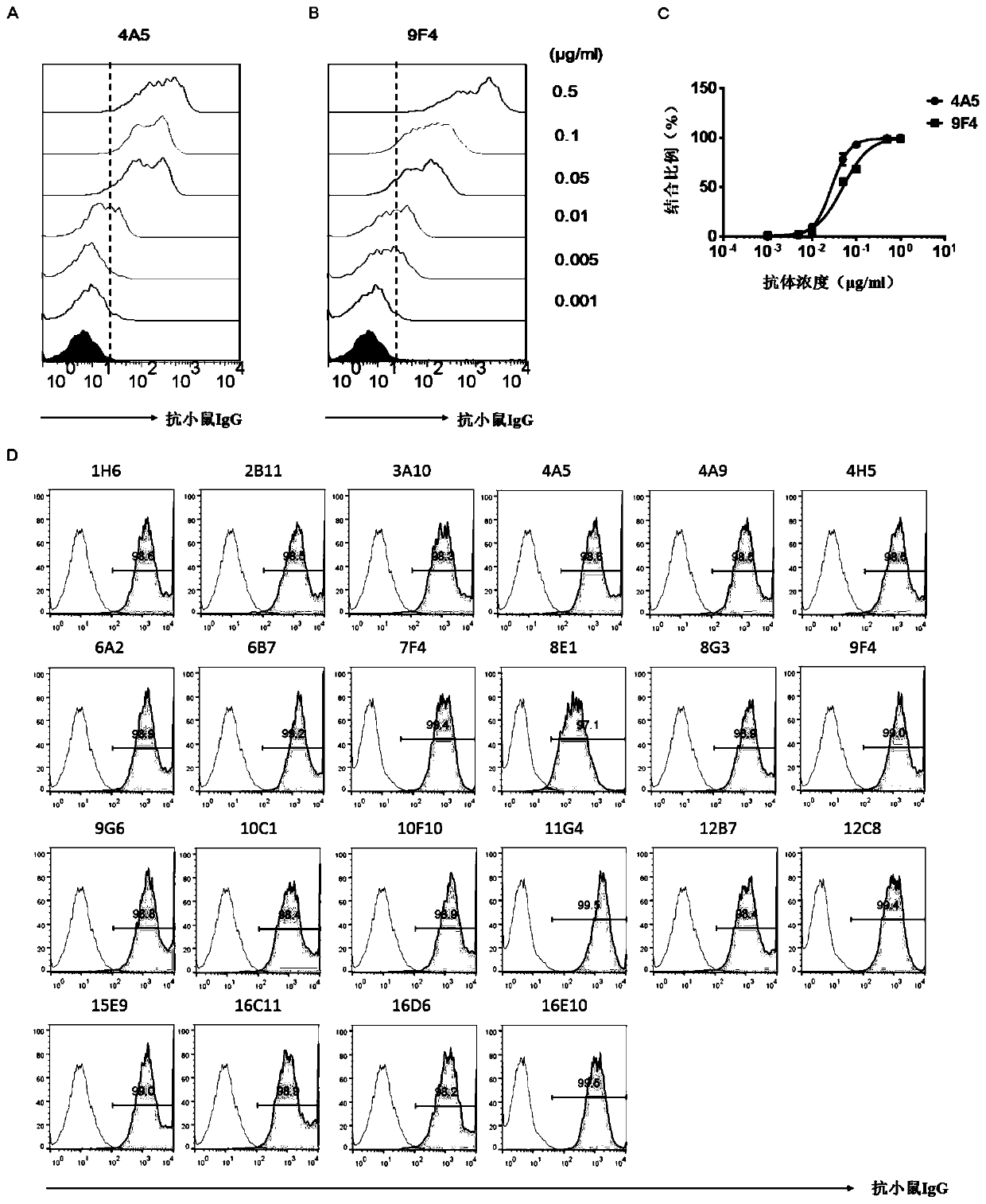
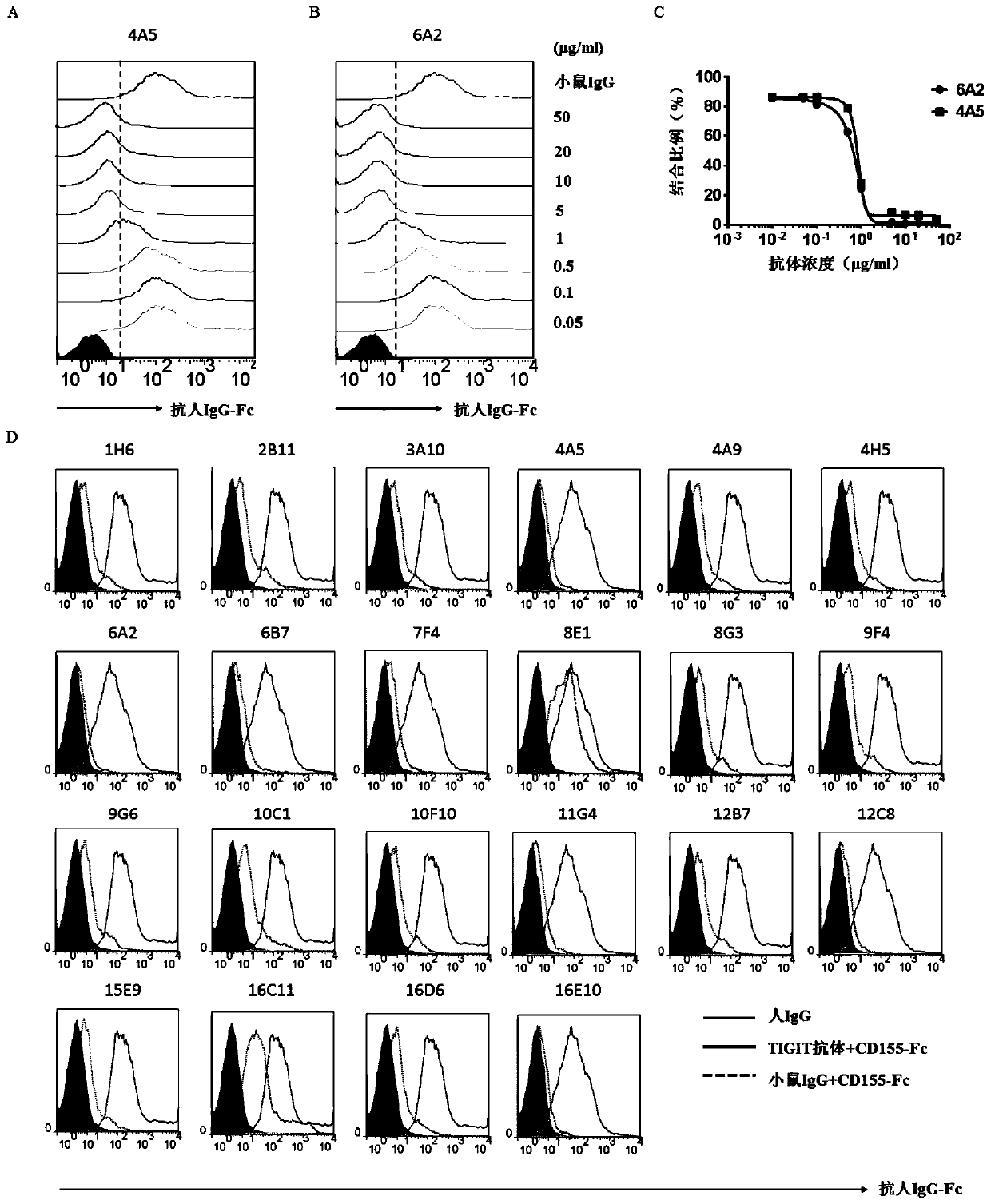
[0160] The immunized mice were immunized three times with purified antigen and complete Freund's adjuvant, and the immune response was detected after bloodletting from the tail vein. The serum was screened by ELISA and flow cytometry, and mice with anti-human TIGIT immunoglobulin were obtained. And for the mouse with the highest anti-TIGIT immunoglobulin, the splenocytes were taken out and fused with the mouse myeloma cell SP2 / 0 cell (ATCC number CRL-1581). The fused hybridoma cells were screened for antibodies to obtain mouse monoclonal antibodies 1H6, 2B11, 3A10, 4A5, 4A9, 4...
Embodiment 2
[0162] Example 2: Antibody binding ability screening
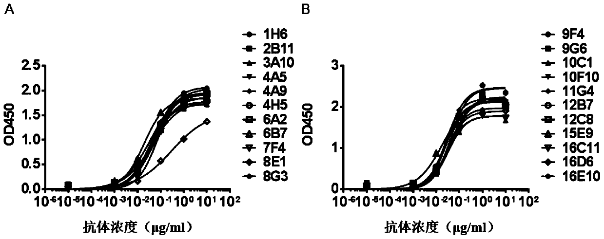
[0163] TIGIT Antibody ELISA Binding Experiment
[0164] ELISA experiments were used to examine the binding properties of TIGIT antibodies. The TIGIT extracellular region Fc fusion protein (TIGIT-Fc) was coated into a 96-well plate, and the signal intensity after the antibody was added was used to determine the binding characteristics of the antibody and TIGIT.
[0165] The TIGIT-Fc fusion protein (amino acid sequence shown in SEQ ID NO: 121) was diluted to 1 μg / ml with PBS buffer, added to a 96-well plate at a volume of 100 μl / well, and left overnight at 4°C. Aspirate the PBS buffer in the 96-well plate, wash the plate 6 times with PBST (pH7.2 PBS containing 0.1% Tween 20) buffer, add 200 μl / well PBS / 10% BSA, and incubate at 37°C for 2 hours to block. Remove the blocking solution, wash the plate 6 times with PBST, add 100 μl / well of the TIGIT antibody to be tested diluted to an appropriate concentration with PBST / 0.05% B...
Embodiment 3
[0170] Embodiment 3: In vitro binding affinity and kinetic experiments
[0171] The Biacore method is a recognized method for objectively detecting the mutual affinity and kinetics of proteins, and the TIGIT antibody of the present invention is analyzed by Biacore T200 to characterize the affinity and binding kinetics.
[0172] The TIGIT extracellular fragment Fc fusion protein (TIGIT-Fc) was covalently linked to the CM5(GE) chip by standard amino coupling method. A series of concentration gradients of TIGIT antibody diluted in PBS was then injected with each cycle and regenerated with 10 mM NaOH solution after injection. Track the antigen-antibody binding kinetics for 3 minutes and track the dissociation kinetics for 10 minutes, use GE's BIAevaluation software to analyze the data obtained with a 1:1 (Langmuir) binding model, and the ka (kon) and kd (koff) determined by this method and KD values are shown in Table 3 below.
[0173] Table 3 TIGIT antibody affinity
[0174]...
PUM
 Abstract
Description
Claims
Application Information
 - R&D
- Intellectual Property
- Life Sciences
- Materials
- Tech Scout
- Unparalleled Data Quality
- Higher Quality Content
- 60% Fewer Hallucinations
Browse by: Latest US Patents, China's latest patents, Technical Efficacy Thesaurus, Application Domain, Technology Topic, Popular Technical Reports.
© 2025 PatSnap. All rights reserved.Legal|Privacy policy|Modern Slavery Act Transparency Statement|Sitemap|About US| Contact US: help@patsnap.com


