
Vascular segmentation method and device and computer device

A perfusion imaging and image segmentation technology, applied in the field of image processing, can solve the problems of blood vessel insufficiency and reducing the accuracy of blood vessel segmentation

Active Publication Date: 2019-03-08
SHANGHAI UNITED IMAGING HEALTHCARE
View PDF9 Cites 14 Cited by
  • Summary
  • Abstract
  • Description
  • Claims
  • Application Information

AI Technical Summary

Problems solved by technology

[0006] The technical problem to be solved by the present invention is to provide a blood vessel segmentation method and device based on perfusion imaging and a computer device in order to overcome the defect that the blood vessel segmentation method in the prior art is likely to cause incomplete blood vessels, thereby seriously reducing the accuracy of blood vessel segmentation

Method used


Image

  • Vascular segmentation method and device and computer device
  • Vascular segmentation method and device and computer device
  • Vascular segmentation method and device and computer device

Examples


Embodiment Construction

[0122] The present invention will be described in detail below in conjunction with the accompanying drawings and specific embodiments. Note that the aspects described below in conjunction with the drawings and specific embodiments are only exemplary, and should not be construed as limiting the protection scope of the present invention.

[0123] The following description is given to enable a person skilled in the art to make and use the invention and incorporate it into a specific application context. Various modifications, and various uses in different applications will be readily apparent to those skilled in the art, and the general principles defined herein may be applied to a wide range of embodiments. Thus, the present invention is not limited to the embodiments given herein but is to be accorded the widest scope consistent with the principles and novel features disclosed herein.

[0124] In the following detailed description, numerous specific details are set forth in or...


PUM


Abstract

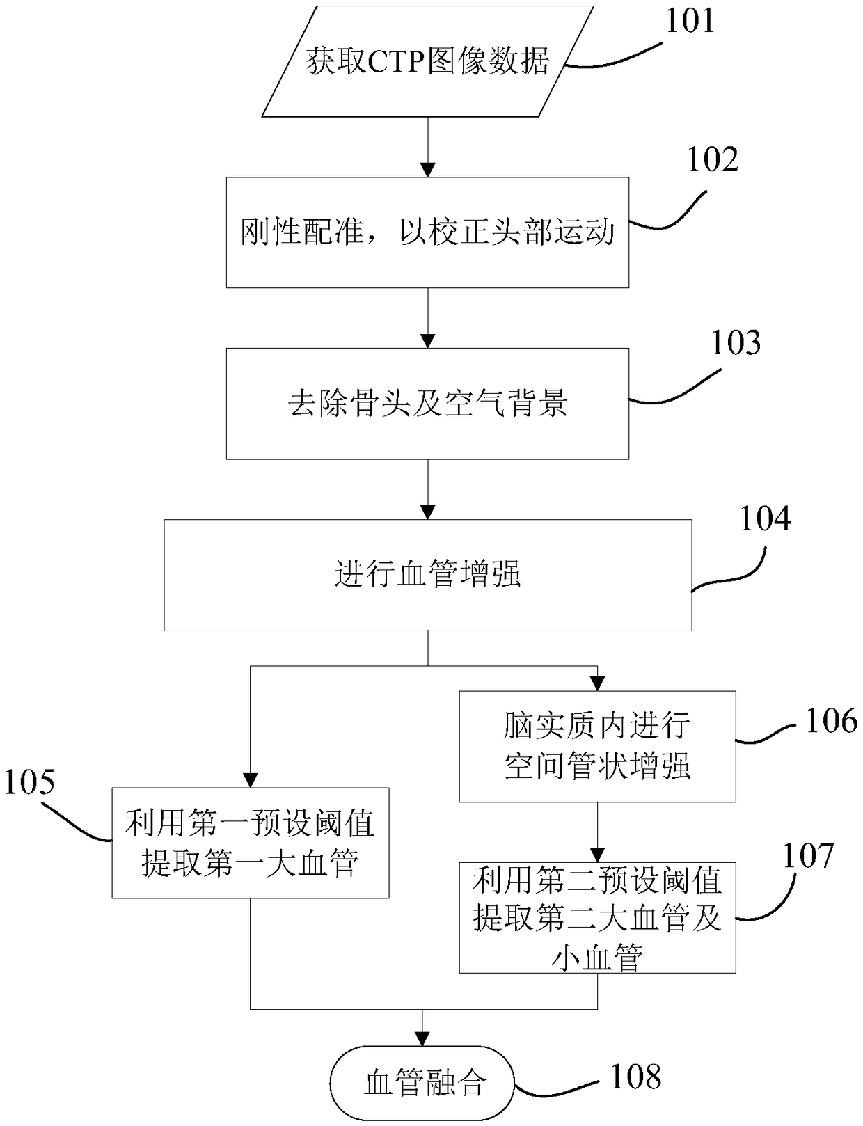
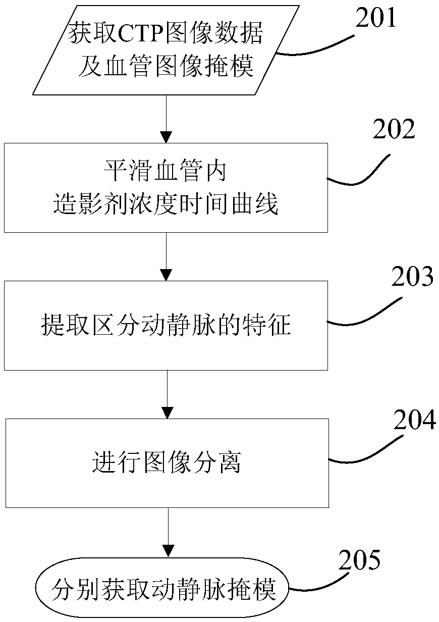
The invention discloses a vascular segmentation method and device and a computer device. The vascular segmentation method includes: acquiring perfusion image data of a selected object; performing vascular reinforcement on the perfusion image data on the basis of change information of concentration of a contrast in a vessel and / or vascular space tubular structural information to obtain vascular reinforcement image data; using a preset threshold to perform image segmentation on the vascular reinforcement image data so as to extract macro-vascular image data and micro-vascular image data; fusingthe macro-vascular image data and the micro-vascular image data to obtain a vascular image mask. Vascular reinforcement is performed through time and space information, such as changes in the contrastin the vessels and tubular structure of the vessels so as to extract macro vessels and micro vessels, the influence of non-rigid deformation portions, such as nasal cavity, upon vascular segmentationis effectively avoided, richer vascular structure can be extracted for clinical diagnosis and reference, and vascular segmentation accuracy is greatly improved.

Description

technical field [0001] The present invention relates to the technical field of image processing, in particular to a blood vessel segmentation method and device based on perfusion imaging and a computer device. Background technique [0002] In recent years, the research and application of perfusion in the diagnosis, prognosis and efficacy evaluation of tumors, especially brain tumors, have become more and more extensive. Perfusion imaging can be used to quantitatively analyze the distribution of microvessels and blood perfusion status in tissues, provide diagnostic basis for abnormal cerebral perfusion diseases (such as stroke, etc.), and provide effective reference for making treatment decisions. [0003] For example, the clinical diagnosis of cerebral apoplexy usually requires the combination of unenhanced CT (Computed Tomography, computerized tomography) and angiography (CT Angiography, CTA) to diagnose cerebral apoplexy, but there is a relatively large radiation dose. CT...

Claims


Application Information

Patent Timeline
Patent Type & Authority Applications(China)
IPC IPC(8): A61B6/03G06T7/136G06K9/62
CPCG06T7/136A61B6/032A61B6/481A61B6/504A61B6/5217G06T2207/10081G06T2207/30101G06F18/23213
Inventor 龙帆李阳
Owner SHANGHAI UNITED IMAGING HEALTHCARE
Learn More