A kind of ocular surface repair film and preparation method thereof

A serosal and mucous layer technology, applied in medical science, tissue regeneration, prostheses, etc., can solve problems such as inappropriate hardness and hinder the wide application of ECM, improve biological safety, avoid secondary damage and shortage of donors , promote the effect of epithelialization

Active Publication Date: 2021-12-03
广州聚明生物科技有限公司
View PDF7 Cites 0 Cited by
  • Summary
  • Abstract
  • Description
  • Claims
  • Application Information

AI Technical Summary

Problems solved by technology

Although these technologies have successfully obtained extracellular matrix (ECM) materials, there are still certain disadvantages when they are used in the repair of ocular surface defects. For example, the hardness of these extracellular matrix materials is not suitable, which seriously hinders the use of ECM in ocular surface repair. widely used

Method used


Image

  • A kind of ocular surface repair film and preparation method thereof

Examples


Embodiment 1

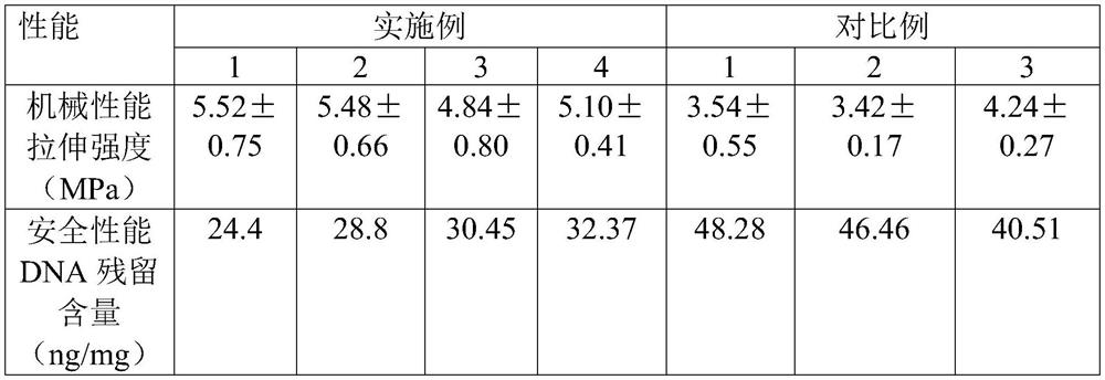
[0026] This embodiment provides a porcine small intestinal submucosa tissue decellularized matrix material and a preparation method thereof. The preparation method includes:

[0027] Step 1. Take the porcine small intestine jejunum section, clean it, cut it radially, and wash it repeatedly with PBS solution (phosphate buffer solution) to remove pollutants.

[0028] Step 2. Physically remove the mucous membrane, using a wooden scraper, carefully and evenly scrape off the mucous membrane layer of the inner layer of the SIS membrane.

[0029] Step 3. Physically remove the sarcolemma and serosa. The wooden scraper and scalpel are used together to remove the sarcolemma and serosa on the outer layer of the SIS membrane.

[0030] Step 4, configuring 0.2% (mass fraction) peracetic acid solution, and then soaking the SIS membrane with the mucous membrane layer, sarcolemma layer and serosa layer removed in 0.2% peracetic acid solution for 7 hours.

[0031] Step 5, taking out the SIS d...

Embodiment 2

[0039] This embodiment provides a porcine small intestinal submucosa tissue decellularized matrix material and a preparation method thereof. The preparation method includes:

[0040] Step 1. The jejunum section of the porcine small intestine was taken, cleaned and cut radially, and washed repeatedly with PBS solution to remove pollutants.

[0041] Step 2. Physically remove the mucous membrane, using a wooden scraper, carefully and evenly scrape off the mucous membrane layer of the inner layer of the SIS membrane.

[0042] Step 3. Physically remove the sarcolemma and serosa. The wooden scraper and scalpel are used together to remove the sarcolemma and serosa on the outer layer of the SIS membrane.

[0043] Step 4, configuring 0.2% (mass fraction) sodium hydroxide solution, and then soaking the SIS membrane with the mucous membrane layer, sarcolemma layer and serosa layer removed in 0.2% sodium hydroxide solution for 7 hours.

[0044] Step 5. Take out the SIS decellularized ma...

Embodiment 3

[0052] This embodiment is a modification example of Embodiment 1, and the main changes are the concentrations of the peracetic acid solution and the sodium hydroxide solution and the soaking time. The concentration of the peracetic acid solution used in this embodiment is 0.1%, the soaking time is 7h, the concentration of the sodium hydroxide solution is 0.5%, and the soaking time is 4h.


PUM


Abstract

The invention relates to an ocular surface repair film and a preparation method thereof. The preparation method comprises: (1) taking pig small intestine jejunum section, cleaning, radially incision, removing mucosal layer, sarcolemma layer and serosa layer to obtain pig small intestine submucosa tissue membrane; The porcine small intestinal submucosa tissue membrane is respectively soaked in peracetic acid solution and sodium hydroxide solution, taken out and washed to obtain the acellular matrix of porcine small intestinal submucosa tissue; (3) soaking the porcine small intestinal submucosa obtained in step (2) with dopamine solution Tissue acellular matrix, the porcine small intestinal submucosa tissue acellular matrix material was obtained. The acellular matrix material of porcine small intestinal submucosa tissue obtained by the preparation method has relatively suitable mechanical hardness, and such mechanical hardness can ensure that the acellular matrix of porcine small intestinal submucosa tissue can be well attached to the ocular surface.

Description

technical field [0001] The invention relates to the technical field of tissue repair materials, in particular to an ocular surface repair membrane and a preparation method thereof, in particular to a porcine small intestinal submucosa tissue decellularized matrix material and a preparation method thereof. Background technique [0002] Ocular surface disease is a common and frequently-occurring disease in ophthalmology, and has a high recurrence rate. It is one of the major blinding diseases in ophthalmology. Examples include chemical burns, superficial ocular surface ulcers, scleral ulcers and perforations, pterygium surgery, and ocular surface trauma. For the treatment of ocular surface epithelial cell defects caused by various accidents, ocular surface reconstruction has the best effect at present, and the commonly used method is autologous or allogeneic ocular surface transplantation. Among them, the ocular surface epithelium is transparent and avascular, located in the ...

Claims


Application Information

Patent Timeline
Patent Type & Authority Patents(China)
IPC IPC(8): A61L27/36A61L27/50
CPCA61L27/3633A61L27/3687A61L27/50A61L2430/16A61L2430/40
Inventor 凌友杨景刘小敏
Owner 广州聚明生物科技有限公司
Why Eureka
Try Eureka