Lesion image generation method, device and computer-readable storage medium

An image generation and image technology, applied in the computer field, can solve problems such as heavy workload, difficulty in obtaining medical images, and affecting technological development, and achieve the effect of reducing requirements, improving acquisition efficiency, and reducing demand

Active Publication Date: 2019-03-26
苏州六莲科技有限公司
View PDF11 Cites 5 Cited by
  • Summary
  • Abstract
  • Description
  • Claims
  • Application Information

AI Technical Summary

Problems solved by technology

But we know that due to the confidentiality of medical information, it is difficult for us to obtain a large number of available medical images, and image segmentation is time-consuming and laborious, which makes the training of image segmentation technology a huge workload, which in turn affects the development of this technology

Method used


Image

  • Lesion image generation method, device and computer-readable storage medium
  • Lesion image generation method, device and computer-readable storage medium
  • Lesion image generation method, device and computer-readable storage medium

Examples


Embodiment Construction

[0023] The technical solutions in the embodiments of the present disclosure will be clearly and completely described below with reference to the accompanying drawings in the embodiments of the present disclosure. Obviously, the described embodiments are only a part of the embodiments of the present disclosure, but not all of the embodiments. The following description of at least one exemplary embodiment is merely illustrative in nature and is in no way intended to limit the disclosure, its application or uses in any way. Based on the embodiments in the present disclosure, all other embodiments obtained by those of ordinary skill in the art without creative efforts shall fall within the protection scope of the present disclosure.

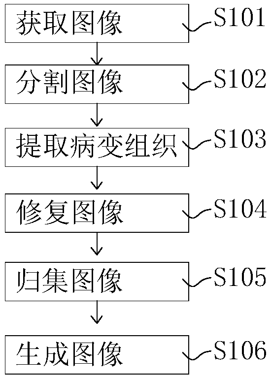
[0024] A method of image segmentation provided by the present disclosure will combine figure 1 describe.

[0025] like figure 1 A schematic flowchart of a method for generating lesion images according to some embodiments is shown. Including steps ...


PUM


Abstract

The invention discloses a Lesion image generation method, device and computer-readable storage medium, which relates to the computer field, in particular to the medical image processing field. The method comprises the following steps: obtaining a lesion medical image; Segmentation of the medical image to obtain a segmented image; Extracting the pathological tissue from the pathological medical image; restoring The medical images of the corresponding organs after the pathological tissues were extracted. grouping The images of pathological tissue extracted from the same organ together to form aset of pathological tissue images. grouping Non-pathological medical images of the same organ into a set of non-pathological medical images; misfusing The image set of pathological tissue and the non-pathological medical image set to generate new pathological medical image of the organ. The invention also provides a lesion image generating device and a computer-readable storage medium. A large numb of lesion medical images are quickly generate by using that original lesion medical images, and the efficiency of obtaining the lesion medical image is greatly improved.

Description

【Technical field】 [0001] The present invention relates to the field of computers, in particular to a method, device and computer-readable storage medium for generating lesion images. 【technical background】 [0002] Digital medical image technology is one of the most important diagnostic methods in modern medicine. With the continuous development and popularization of digital medical image diagnostic technology, doctors have higher and higher requirements for medical image diagnosis. For 3D model diagrams, further quantitative analysis, simulation, and combined use with AR / VR are required. 3D reconstruction of medical images is an important technology for converting 2D images into 3D models. If quantitative analysis is required, different organs / tissues need to be segmented and reconstructed separately before quantitative analysis or simulation of organs or tissues can be performed. [0003] Since medical images conforming to the DICOM3.0 standard are two-dimensional images ...

Claims


Application Information

Patent Timeline
Patent Type & Authority Applications(China)
IPC IPC(8): G06T7/00G06T7/11G06T5/50G16H30/40
CPCG06T5/50G06T7/0012G06T7/11G16H30/40G06T2207/20221
Inventor 钟亚玉廖珍琪张健莫康信其他发明人请求不公开姓名
Owner 苏州六莲科技有限公司
Why Eureka
Try Eureka