A retinal fundus image segmentation method based on a deep full convolutional neural network

A convolutional neural network and fundus image technology, applied in the field of medical image processing, can solve the problems of long time required, relatively sensitive selection, complex processing process, etc., and achieve the effect of ensuring segmentation accuracy, fast segmentation speed, and high processing effect.

Active Publication Date: 2019-04-09
NANJING UNIV OF AERONAUTICS & ASTRONAUTICS
View PDF4 Cites 21 Cited by
  • Summary
  • Abstract
  • Description
  • Claims
  • Application Information

AI Technical Summary

Problems solved by technology

[0003] The optic cup and optic disc segmentation methods of retinal fundus images can be roughly divided into three categories: template-based methods, deformation model-based methods, and pixel-based classification methods. The first two methods are mainly based on the edge features of the optic cup and optic disc. The implementation is very dependent on the edge difference between the optic cup and optic disc edge and other structural regions. When there are confusing lesions, the segmentation algorithm is not effective, and the method based on the deformation model is relatively sensitive to the selection of the initial point. For a good initialization Point selection is relatively difficult
The method based on pixel classification is greatly limited by the high number of pixels in high-resolution images, and it is very difficult to achieve model optimization at the pixel level
At the same time, the processing process of the above segmentation method is relatively complicated, and the time required for image segmentation is relatively long.

Method used


Image

  • A retinal fundus image segmentation method based on a deep full convolutional neural network
  • A retinal fundus image segmentation method based on a deep full convolutional neural network
  • A retinal fundus image segmentation method based on a deep full convolutional neural network

Examples


Embodiment 1

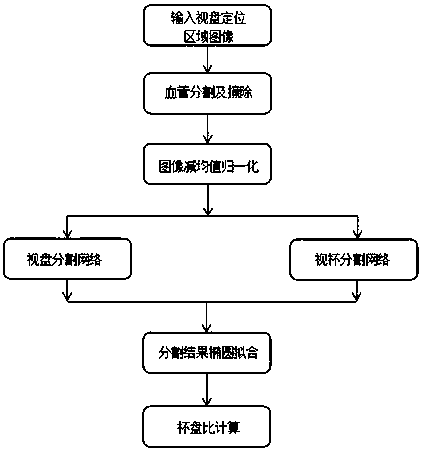
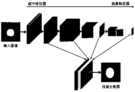
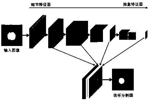
[0024] Embodiment 1: The present invention provides a method for segmenting retinal fundus images based on a deep full convolutional neural network. First, based on an existing algorithm, the optic disc region of the fundus image is located and extracted, and then the optic disc positioning region image is used as a deep full convolutional neural network. The input of the network is then used to predict the pixels in the input image using a deep fully convolutional neural network, and finally the corresponding cup-to-disk ratio is calculated through the obtained optic disc and optic cup segmentation results, such as figure 1 shown.

[0025] The method and technical effect of the present invention will be described below through specific examples.

[0026] Step 1: Use the fundus map dataset ORIGA as the training and test retinal fundus image set, which has a total of 650 left and right eye images of different objects. Among them, 325 images are used as training samples, and th...


PUM


Abstract

The invention discloses a retinal fundus image segmentation method based on a deep full convolutional neural network, and the method comprises the steps: selecting a training set and a test set, carrying out the extraction of a retinal fundus image, obtaining an optic disc positioning area image, and carrying out the blood vessel removal operation; Constructing a deep full convolutional neural network, taking the image of the optic disc positioning area as the input of the deep full convolutional neural network, performing optic disc segmentation model training on the training set based on thetrained weight parameter as an initial value to finely adjust model parameters, and performing parameter fine adjustment on the optic disc segmentation model on the basis; Using the trained visual cup segmentation model to perform visual cup and visual disc segmentation on the test set, performing ellipse fitting on a final segmentation result, and calculating a vertical cup-visual disc ratio according to a segmentation boundary of the visual cup and the visual disc. Automatic segmentation of the optic disc and the optic cup of the retinal fundus image is realized, the precision is high, andthe speed is high.

Description

technical field [0001] The invention relates to a retinal fundus image segmentation method based on a deep full convolutional neural network, and belongs to the field of medical image processing. Background technique [0002] The cup-to-disc vertical ratio is one of the important basis for doctors to judge various fundus diseases. In current clinical practice, the cup-to-disc ratio is mostly measured and calculated manually by doctors, but manual measurement is not only time-consuming and laborious, but also the measurement results of different doctors. There is a certain subjectivity, so it is not suitable for large-scale disease screening. Therefore, the calculation of cup-to-disk ratio for automatic cup-to-disk segmentation has attracted more and more attention. [0003] The optic cup and optic disc segmentation methods of retinal fundus images can be roughly divided into three categories: template-based methods, deformation model-based methods, and pixel classification-...

Claims


Application Information

Patent Timeline
Patent Type & Authority Applications(China)
IPC IPC(8): G06T7/11
CPCG06T7/11G06T2207/20081G06T2207/20084G06T2207/30041
Inventor 万程牛笛周鹏彭琦华骁
Owner NANJING UNIV OF AERONAUTICS & ASTRONAUTICS
Why Eureka
Try Eureka