Cell nucleus segmentation method and device

A cell nucleus and cell technology, applied in the field of medical image processing, can solve the problems of poor applicability of multiple types of histopathological images

Pending Publication Date: 2020-06-30
UNIV OF SCI & TECH OF CHINA +1
View PDF7 Cites 2 Cited by
  • Summary
  • Abstract
  • Description
  • Claims
  • Application Information

AI Technical Summary

Problems solved by technology

[0005] This application provides a cell nucleus segmentation method and device, with the purpose of solving the problem of poor applicability of existing segmentation methods to multi-category histopathological images

Method used


Image

  • Cell nucleus segmentation method and device
  • Cell nucleus segmentation method and device
  • Cell nucleus segmentation method and device

Examples


Embodiment Construction

[0067] The following will clearly and completely describe the technical solutions in the embodiments of the application with reference to the drawings in the embodiments of the application. Apparently, the described embodiments are only some of the embodiments of the application, not all of them. Based on the embodiments in this application, all other embodiments obtained by persons of ordinary skill in the art without making creative efforts belong to the scope of protection of this application.

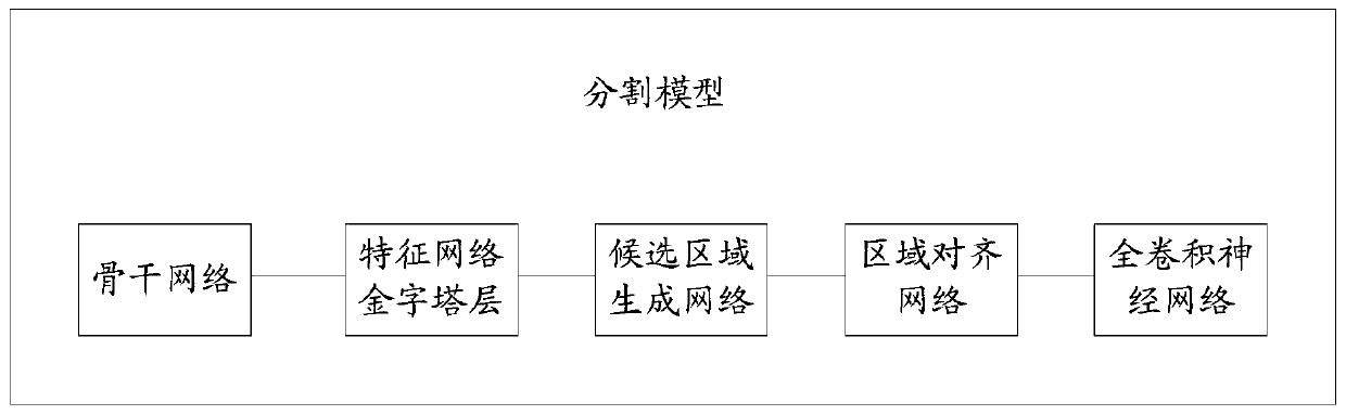
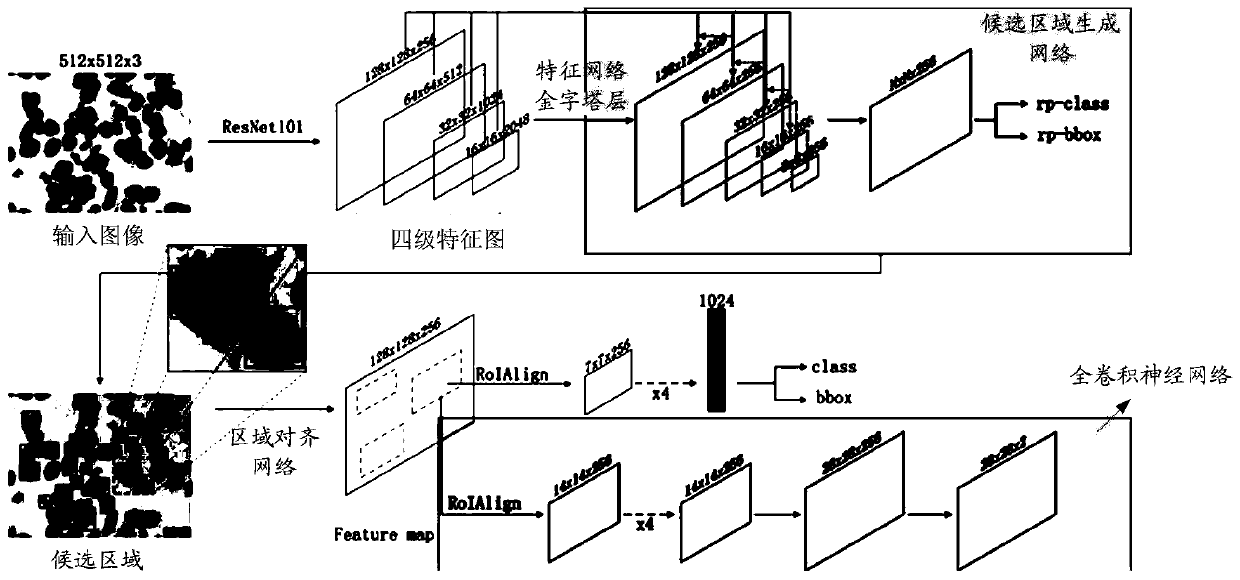
[0068] figure 1 A schematic structural diagram of a segmentation model provided for the embodiment of the present application, including: a backbone network, a feature network pyramid layer, a candidate region generation network, a region alignment network, and a preset full convolutional neural network, which are sequentially connected.

[0069] Among them, the backbone network is obtained by removing the global pooling layer and the fully connected layer of the ResNet101 model. S...


PUM


Abstract

The invention provides a cell nucleus segmentation method and device, and the method comprises the steps: inputting a to-be-segmented image into a trained segmentation model under the condition that the resolution of the to-be-segmented image is not greater than a preset resolution, and obtaining a segmentation result of a cell nucleus in the to-be-segmented image, wherein the preset resolution isdetermined by the fixed input resolution of the trained segmentation model, and the trained segmentation model is obtained by respectively training preset segmentation models by adopting different types of training samples; training a preset segmentation model by taking a preset natural scene image as a training sample to obtain a first segmentation model; training the first segmentation model bytaking the multi-class tissue pathological images corresponding to the various tissues as training samples to obtain a second segmentation model; and finally, training the second segmentation model by taking the multi-class histopathological image of the required tissue as a training sample to obtain a trained segmentation model. The applicability of the segmentation method is improved.

Description

technical field [0001] The present application relates to the field of medical image processing, in particular to a cell nucleus segmentation method and device. Background technique [0002] Pathological diagnosis is the "gold standard" for diagnosis of various diseases. The pathological diagnosis mode is to combine the morphology of tissue cells with clinical, imaging and inspection for comprehensive analysis and judgment to make a final diagnosis of the disease. The analysis of pathological tissue cell morphology is to check the cell morphology characteristics of a given tissue by H&E staining, IHC (immunohistochemistry), molecular pathology, etc. (for the convenience of description, different imaging methods, such as brightfield (brightfield) and fluorescence (fluorescence, different cell staining methods, such as H&E staining and other images obtained by imaging a given tissue, called multi-category histopathological images), is the core of pathological diagnosis techno...

Claims


Application Information

Patent Timeline
Patent Type & Authority Applications(China)
IPC IPC(8): G06T7/00G06T7/11G06T7/136
CPCG06T7/0012G06T7/11G06T7/136G06T2207/10056G06T2207/20081G06T2207/20084G06T2207/20016G06T2207/30024
Inventor 安虹吕国锋韩文廷陈俊仕石军王朝晖文可李秀林
Owner UNIV OF SCI & TECH OF CHINA
Why Eureka
Try Eureka