Closed medical imaging diagnostic instrument for radiology department

A medical imaging and closed technology, applied in the directions of radiological diagnosis instruments, diagnosis, patient positioning for diagnosis, etc., can solve problems such as unfavorable diagnosis work and patients are easily disturbed by the outside world, and achieve sufficient diagnosis and good effect. Effect

Pending Publication Date: 2019-04-12
程显峰
View PDF0 Cites 2 Cited by
  • Summary
  • Abstract
  • Description
  • Claims
  • Application Information

AI Technical Summary

Problems solved by technology

[0004] Existing medical imaging diagnostic instruments are generally open, so that patients are susceptible to external interference during diagnosis, and external personnel may also be affected by internal diagnostic devices, which is not conducive to the diagnosis work

Method used


Image

  • Closed medical imaging diagnostic instrument for radiology department
  • Closed medical imaging diagnostic instrument for radiology department
  • Closed medical imaging diagnostic instrument for radiology department

Examples


Embodiment 1

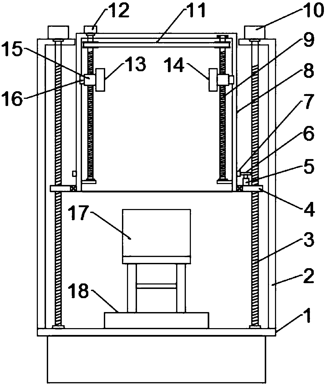
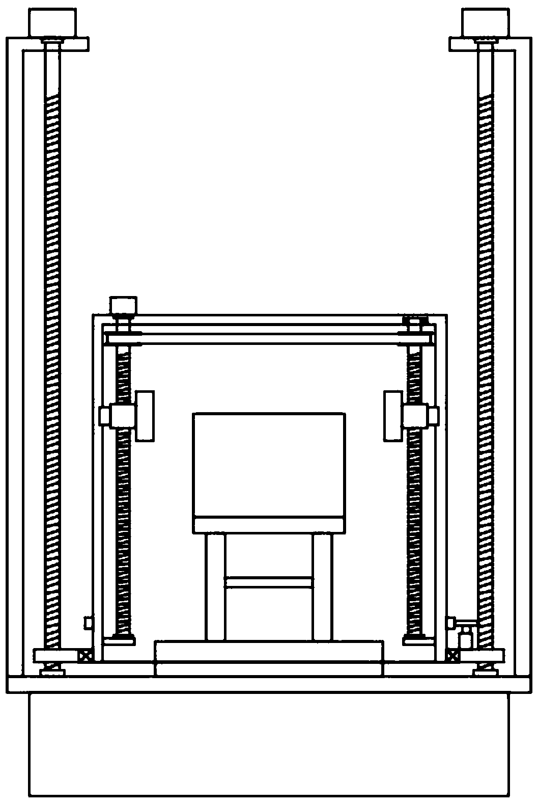
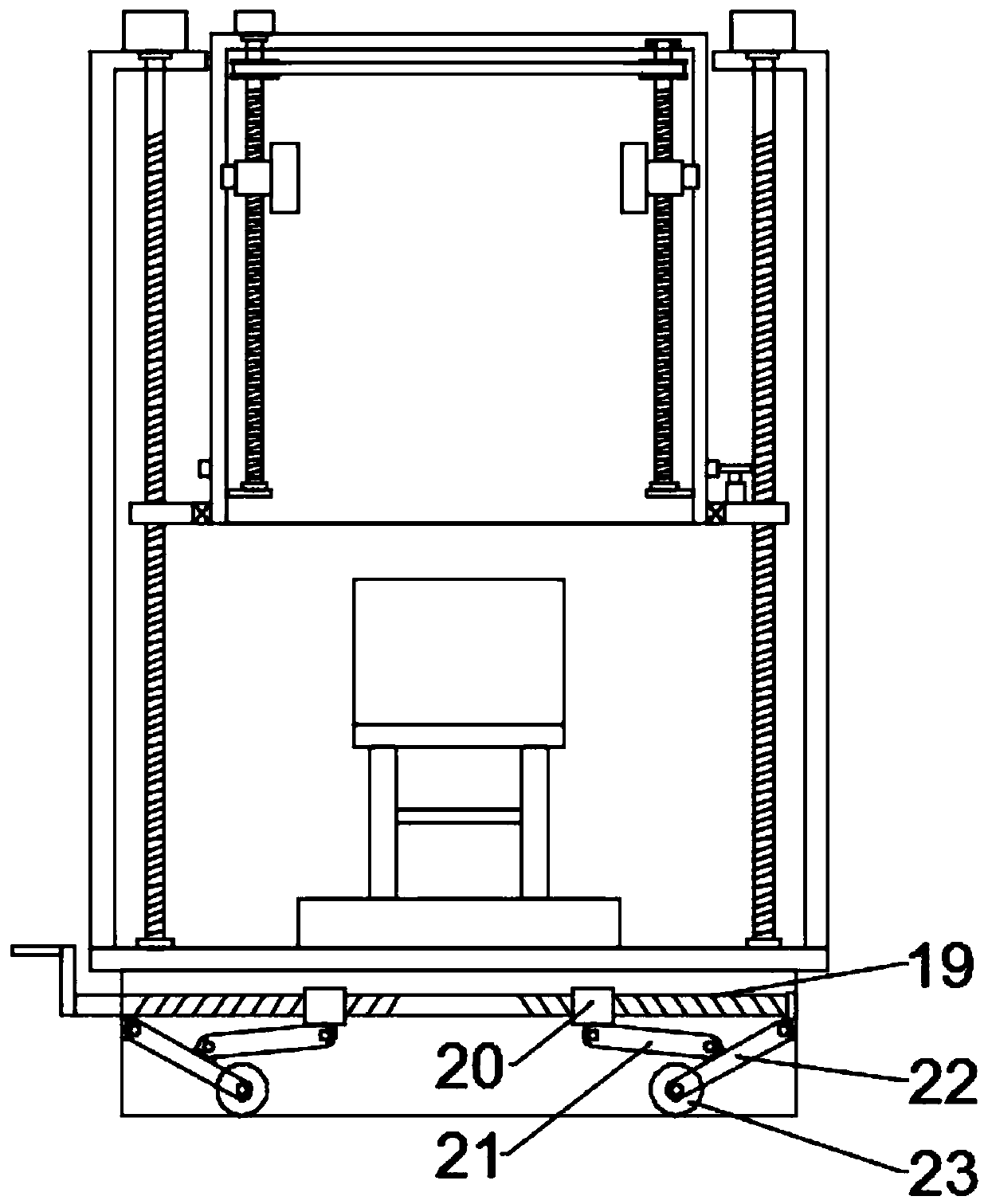
[0026] see Figure 1~2 , in an embodiment of the present invention, a closed medical imaging diagnostic instrument for radiology, including a substrate 1, an imaging device 13 and a scanning device 14, the substrate 1 is installed on a support base, and a workbench 18 is provided on the upper surface of the substrate 1 , a seat 17 is installed on the workbench 18, an L-shaped plate 2 is fixed on both sides of the top of the base plate 1, and a first threaded rod 3 is installed on the L-shaped plate 2 through bearing rotation, and the top of the first threaded rod 3 passes through a coupling Fixedly connected with the output shaft of the first motor 10, the first motor 10 is mounted on the L-shaped plate 2 by screws, and a lifting plate 4 is connected between the two first threaded rods 3, and the first threaded rod 3 and the lifting plate 4 threaded connection, the lifting plate 4 is equipped with a closing cover 8, the lifting plate 4 is provided with a mounting hole for inst...

Embodiment 2

[0028] see Figure 1~3 , in an embodiment of the present invention, a closed medical imaging diagnostic instrument for radiology, including a substrate 1, an imaging device 13 and a scanning device 14, the substrate 1 is installed on a support base, and a workbench 18 is provided on the upper surface of the substrate 1 , a seat 17 is installed on the workbench 18, an L-shaped plate 2 is fixed on both sides of the top of the base plate 1, and a first threaded rod 3 is installed on the L-shaped plate 2 through bearing rotation, and the top of the first threaded rod 3 passes through a coupling Fixedly connected with the output shaft of the first motor 10, the first motor 10 is mounted on the L-shaped plate 2 by screws, and a lifting plate 4 is connected between the two first threaded rods 3, and the first threaded rod 3 and the lifting plate 4 threaded connection, the lifting plate 4 is equipped with a closing cover 8, the lifting plate 4 is provided with a mounting hole for inst...


PUM


Abstract

The invention discloses a closed medical imaging diagnostic instrument for radiology department. The closed medical imaging diagnostic instrument for the radiology department comprises a base plate, an imaging device and a scanning device; the base plate is mounted on a support base; a table is arranged on an upper surface of the base plate; L-shaped boards are fixed on both sides of the top of the base plate; first threaded rods are rotatably mounted on the L-shaped boards via bearings; a lifting board is connected between the two first threaded rods; a closing cover is mounted on the liftingboard; second threaded rods are rotatably mounted on both sides of an inner cavity of the closing cover via bearings, and are sleeved with movable blocks; and the imaging device and the scanning device are respectively mounted on the two movable blocks. Being arranged with a first motor, the first threaded rods, the closing cover, a third motor and the second threaded rods, closed space is formedfor the closed medical imaging diagnostic instrument for the radiology department, thereby ensuring smooth and safe performance of diagnosis; moreover, the closed medical imaging diagnostic instrument is also capable of scanning the body of a patient step by step from top to bottom so as to realize fuller diagnosis. In addition, a second motor, a gear and an external gear ring are arranged, so that, 360-degree non-dead-angle omni-directional scanning diagnosis can be realized, thereby achieving better diagnosis effects.

Description

technical field [0001] The invention relates to the technical field of medical equipment, in particular to a closed medical imaging diagnostic instrument for radiology. Background technique [0002] The Department of Radiology is an important auxiliary examination department in a hospital. In the construction of a modern hospital, the Department of Radiology is a department that integrates inspection, diagnosis, and treatment. Many diseases in various clinical departments must be checked by radiology equipment to achieve a clear diagnosis and auxiliary diagnosis. The equipment in the radiology department generally includes ordinary X-ray filming machine, computerized radiography system (CR), direct digital radiography system (DR), computerized tomography (CT), nuclear magnetic resonance (MRI), digital subtraction angiography Angiography system (DSA), etc. [0003] Medical imaging refers to the technology and processing process of obtaining internal tissue images of the huma...

Claims


Application Information

Patent Timeline
Patent Type & Authority Applications(China)
IPC IPC(8): A61B6/00A61B6/04
CPCA61B6/44A61B6/0478
Inventor 程显峰
Owner 程显峰
Why Eureka
Try Eureka