
A breast cancer Ki67/ER/PR nuclear staining cell counting method based on staining separation

A technology for cell counting and breast cancer, applied in computing, image analysis, image data processing, etc., can solve the problems of time-consuming and laborious, subjectivity error of accuracy, etc. Effect

Active Publication Date: 2019-05-03
SHANDONG COMP SCI CENTNAT SUPERCOMP CENT IN JINAN
View PDF6 Cites 12 Cited by
  • Summary
  • Abstract
  • Description
  • Claims
  • Application Information

AI Technical Summary

Problems solved by technology

This manual observation method is time-consuming and laborious, and there is also a large subjective error in accuracy.

Method used


Image

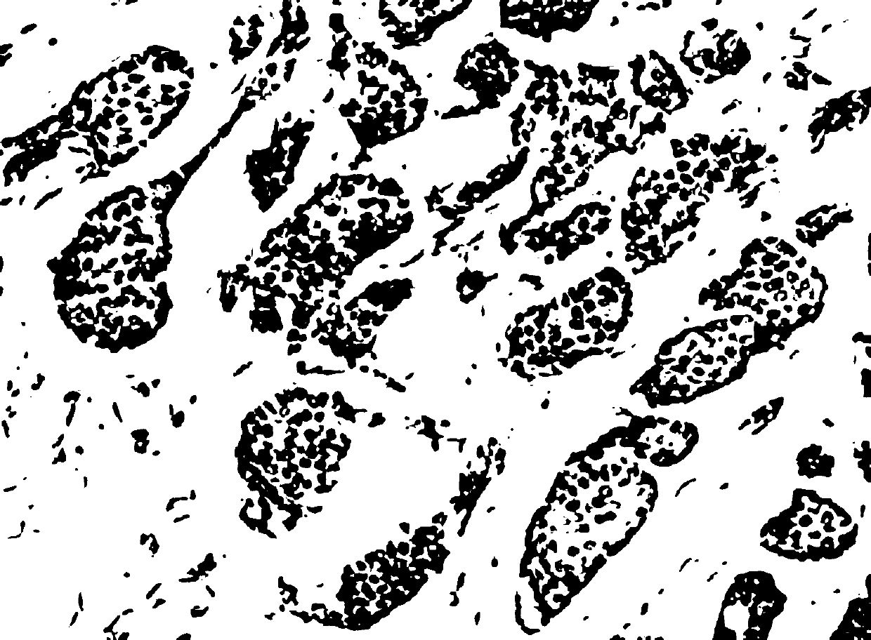
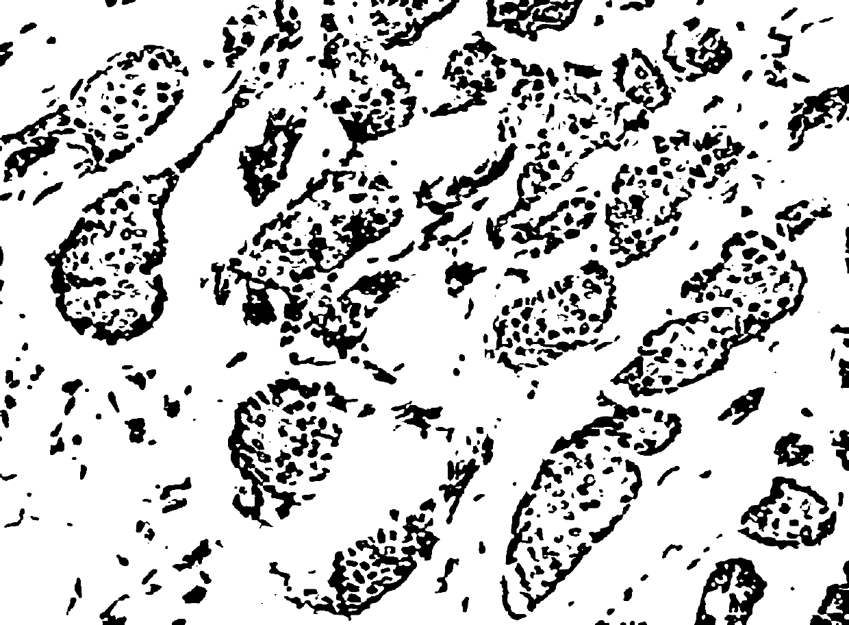
  • A breast cancer Ki67/ER/PR nuclear staining cell counting method based on staining separation
  • A breast cancer Ki67/ER/PR nuclear staining cell counting method based on staining separation
  • A breast cancer Ki67/ER/PR nuclear staining cell counting method based on staining separation

Examples


Embodiment Construction

[0030] The present invention will be further described below in conjunction with the accompanying drawings and embodiments.

[0031] The image data targeted by the present invention are nuclear stained digital pathological sections of breast cancer, including three positive expressions of ER, PR and Ki67. Among them, Ki67 needs to calculate the number of positive and negative cells in the current slice and the probability of each accounting for the total number of cells. ER / PR needs to calculate the number of strong positive, moderate positive, weak positive and negative cells in the current slice, and the respective proportions of the total number of cells. The probability of the number of cells. Nuclear-stained breast cancer pathology sections highlight the color of nuclei by immunohistochemistry (IHC). Immunohistochemistry uses a chemical reaction to develop the chromogenic agent of the labeled antibody, using H staining agent and DAB chromogenic agent. For breast cancer ...


PUM


Abstract

The invention discloses a breast cancer Ki67 / ER / PR nuclear staining cell counting method based on staining separation. The method comprises the steps that firstly, a breast cancer pathological sectionimage is dyed and separated, an image IH based on an H coloring agent and an image IDAB based on a DAB color developing agent are obtained, then the influence of intercellular cytoplasm is eliminatedthrough filtering, and an image IH, filterd, IDAB and filterd are obtained; secondly, counting the negative cells according to IH and filterd, counting strong positive, middle positive and weak positive cells according to IDAB, filterd and the brightness map VDAB of the IH and filterd, and finally obtaining the number of the negative cells, the number of the positive cells and the number of the positive cells (divided into strong positive, middle positive and weak positive) and the probability of occupying the total number of the cells. According to the breast cancer nuclear staining cell counting method based on staining separation, cell counting can be rapidly and effectively achieved, doctors are assisted in work, the workload of the doctors is reduced, and meanwhile the calculation accuracy is guaranteed.

Description

technical field [0001] The invention relates to a method for counting breast cancer Ki67 / ER / PR nuclear staining cells, more specifically, a method for counting breast cancer Ki67 / ER / PR nuclear staining cells based on staining separation. Background technique [0002] According to the national cancer data compiled by the National Cancer Center and the Bureau of Disease Control and Prevention of the Ministry of Health in 2018, the incidence of breast cancer has ranked first in the incidence of female malignant tumors in the country for two consecutive years. The global incidence of breast cancer in women has been on the rise since the late 1970s. China is not a country with a high incidence of breast cancer, but it should not be optimistic. In recent years, the growth rate of the incidence of breast cancer in my country is 1 to 2 percentage points higher than that of countries with a high incidence. For primary breast cancer, immunohistochemical typing is performed internatio...

Claims


Application Information

Patent Timeline
IPC IPC(8): G06T7/00G06T7/155G06T5/40
Inventor 葛菁李娜潘景山赵志刚牛春阳
Owner SHANDONG COMP SCI CENTNAT SUPERCOMP CENT IN JINAN
Learn More