A retinal image segmentation algorithm based on a residual error U-NET network

An image segmentation and retinal technology, applied in image analysis, image enhancement, image data processing, etc., can solve the complex and difficult problems of automatic blood vessel segmentation, achieve the effect of improving visual effects and reducing computational complexity

Inactive Publication Date: 2019-05-07
HARBIN UNIV OF SCI & TECH
View PDF4 Cites 11 Cited by
  • Summary
  • Abstract
  • Description
  • Claims
  • Application Information

AI Technical Summary

Problems solved by technology

In addition, due to the existence of vessel intersections, branches, and centerline reflections, it is also difficult to accurately segment blood v...

Method used


Image

  • A retinal image segmentation algorithm based on a residual error U-NET network
  • A retinal image segmentation algorithm based on a residual error U-NET network
  • A retinal image segmentation algorithm based on a residual error U-NET network

Examples


Embodiment Construction

[0027] The following will clearly and completely describe the technical solutions in the embodiments of the present invention with reference to the accompanying drawings in the embodiments of the present invention. Obviously, the described embodiments are only some, not all, embodiments of the present invention. Based on the embodiments of the present invention, all other embodiments obtained by persons of ordinary skill in the art without making creative efforts belong to the protection scope of the present invention.

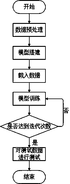
[0028] see figure 1 , the present invention provides a technical solution: a retinal image segmentation method based on the residual U-NET network, comprising the following steps:

[0029] A. Download the color fundus retinal image, and perform sample expansion on the downloaded image;

[0030] B. Preprocessing the original diabetic retinal image;

[0031] C. A residual U-NET network improved by adding a residual structure on the U-NET network;

[0032] D. ...


PUM


Abstract

The invention discloses a retinal image segmentation algorithm based on a residual error U-NET network, which comprises the following steps: A, downloading a color fundus retina image, and carrying out sample expansion on the downloaded image; B, preprocessing the original image; C, obtaining an Improved residual U-NET network by adding a residual structure to the U-NET network; D, taking the processed retina image as input, and carrying out pre-training on the training sample to obtain Initial parameters of the residual U-NET network model; E, Segmenting test samples using a trained residualU-NET neural network model to obtain a final retinal vessel image segmentation map; the residual U-NET neural network model iss adopted to segment the diabetic retina image, the processing effect isgood, the method can be widely applied to the field of diabetic retina diagnosis, and powerful theoretical and technical support is provided for pathological diagnosis of the diabetic retina image.

Description

technical field [0001] The invention relates to the technical field of retinal image processing, in particular to a diabetic retinal image segmentation algorithm based on a residual U-NET network. Background technique [0002] In clinical practice, color fundus images taken by ophthalmoscope are often used as the basis for screening, diagnosis and analysis of related diseases. Therefore, blood vessel segmentation of fundus images has become a prerequisite for quantitative analysis of diseases. For fundus image blood vessel segmentation, the manual segmentation method is very dependent on the operator's experience and technology, and often has shortcomings such as strong subjectivity, low repeatability, high labor intensity, and low efficiency. It is very important in application. Especially with the development of computer-aided diagnosis systems for ophthalmic diseases, the automatic segmentation of fundus blood vessels has been recognized as a very critical and challengin...

Claims


Application Information

Patent Timeline
IPC IPC(8): G06T7/11G06N3/04G06N3/08G06T5/00
Inventor 高俊山魏传雪邓立为
Owner HARBIN UNIV OF SCI & TECH
Why Eureka
Try Eureka