
Positioning method and device for highlight focus area, computer equipment and storage medium

A region and lesion technology, applied in the field of computer equipment, storage media, devices, and positioning methods for highlighted lesion areas, can solve problems such as inaccurate positioning, easy adhesion of highlighted glands in lesion areas, etc.

Active Publication Date: 2019-05-10
SHANGHAI UNITED IMAGING HEALTHCARE
View PDF8 Cites 12 Cited by
  • Summary
  • Abstract
  • Description
  • Claims
  • Application Information

AI Technical Summary

Problems solved by technology

The traditional positioning method for these highlighted areas in mammary images is generally for two-dimensional mammography image processing, and the highlighted area is segmented by simple region growing, but the lesion area is easy to adhere to other highlighted glands, resulting in inaccurate positioning

Method used


Image

  • Positioning method and device for highlight focus area, computer equipment and storage medium
  • Positioning method and device for highlight focus area, computer equipment and storage medium
  • Positioning method and device for highlight focus area, computer equipment and storage medium

Examples


Embodiment Construction

[0038] In order to make the purpose, technical solution and advantages of the present application clearer, the present application will be further described in detail below in conjunction with the accompanying drawings and embodiments. It should be understood that the specific embodiments described here are only used to explain the present application, and are not intended to limit the present application.

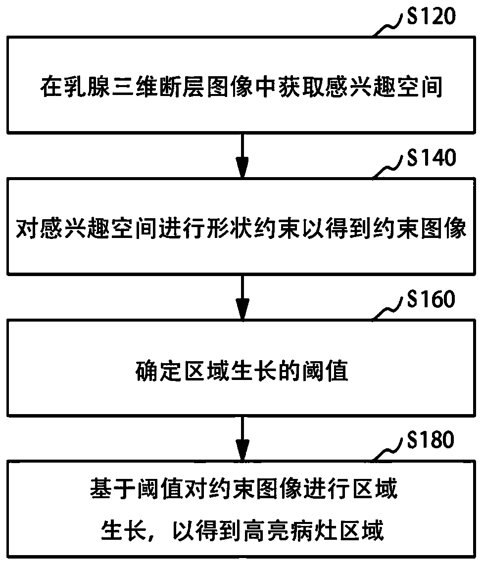
[0039] figure 1 It is a schematic flow chart of the positioning method of the highlighted lesion area in an embodiment, such as figure 1 As shown, a localization method of highlighted lesion area is applied to three-dimensional breast tomographic images, including:

[0040] Step S120: Obtain a space of interest in the three-dimensional tomographic image of the breast.

[0041] Specifically, the above-mentioned highlighted area can generally be a lesion area in a three-dimensional breast tomography image (Digital Breast Tomsynthesis, referred to as DBT), and the lesion ar...


PUM


Abstract

The invention relates to a positioning method and device for a highlight focus area, computer equipment and a storage medium, and the positioning method for the highlight focus area is applied to a mammary gland three-dimensional tomography image and comprises the steps: obtaining an interested space in the mammary gland three-dimensional tomography image; performing shape constraint on the spaceof interest to obtain a constrained image; determining a threshold value of regional growth; and carrying out region growth on the constrained image based on the threshold value to obtain a highlightfocus region. According to the positioning method of the highlight focus area, after the interested space is selected in the mammary gland three-dimensional tomographic image, shape constraint is conducted on the interested space to isolate glands around the highlight focus area, the appropriate area growth threshold value is determined, and therefore segmentation of the highlight focus area is achieved, and many inappropriate threshold values are removed in the positioning process. According to the positioning method, the processing speed is increased, and the segmentation accuracy is improved.

Description

technical field [0001] The invention relates to the field of medical devices, in particular to a positioning method, device, computer equipment and storage medium for a highlighted lesion area. Background technique [0002] When detecting breast tissue, it is often necessary to locate highlighted areas such as lumps and lesions in breast images. The traditional positioning method for these highlighted areas in mammary images is generally for two-dimensional mammography image processing, and the highlighted area is segmented by simple region growing, but the lesion area is easy to adhere to other highlighted glands, resulting in inaccurate positioning . Contents of the invention [0003] Based on this, it is necessary to address the above technical problems and provide a method, device, computer equipment and storage medium for locating the highlighted lesion area, which can locate the highlighted lesion area according to the three-dimensional tomographic image of the brea...

Claims


Application Information

Patent Timeline
IPC IPC(8): G06K9/32G06K9/38G06T7/11G06T7/136
Inventor 吴叶芬
Owner SHANGHAI UNITED IMAGING HEALTHCARE
Learn More