Liver tumor automatic accurate robust segmentation method in CT image

A technology for liver tumors and CT images, applied in instruments, character and pattern recognition, computer components, etc., can solve the uneven gray scale of liver tumors, the inability to obtain liver tumor boundaries, and difficulty in adapting to the complexity and diversity of liver CT images sexual issues

Active Publication Date: 2019-05-14
HUNAN UNIV OF SCI & TECH
View PDF3 Cites 9 Cited by
  • Summary
  • Abstract
  • Description
  • Claims
  • Application Information

AI Technical Summary

Problems solved by technology

These methods only use the underlying image data for segmentation, without combining high-level prior knowledge, and are usually difficult to adapt to the complexity and diversity of liver CT images
The supervised method mainly refers to the method of using image feature prior combined with machine learning for segmentation. Although this type of method can effectively distinguish tumor tissue from normal liver parenchyma by increasing training samples, it can solve the problems of various shapes and uneven gray levels of liver tumors. However, accurate liver tumor boundaries cannot be obtained

Method used


Image

  • Liver tumor automatic accurate robust segmentation method in CT image
  • Liver tumor automatic accurate robust segmentation method in CT image
  • Liver tumor automatic accurate robust segmentation method in CT image

Examples


Embodiment 1

[0057] An automatic accurate and robust segmentation method for liver tumors in CT images, the specific implementation steps are as follows:

[0058] (1) Using sparse shape combination to preprocess the original CT image to obtain the liver region; figure 1 (a)~ figure 1 (c) is three original CT images, figure 1 (d)~ figure 1 (f) is the result of preprocessing it using the method of this embodiment;

[0059] (2) Using the image superpixel segmentation method based on LI-SLIC to perform multi-level iterative segmentation of the liver area, divide the areas with consistent gray scale and texture in the liver into the same superpixel, and obtain the distance between the liver tumor and the normal liver parenchyma Boundary, the superpixel segmentation result is denoted as S i (i=1,2,...,n), where n is the number of superpixels;

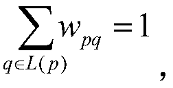
[0060] In the (2) step, the image superpixel segmentation method based on LI-SLIC specifically includes:

[0061] (I) Divide the original image int...


PUM


Abstract

The invention discloses a liver tumor automatic accurate robust segmentation method in a CT image, and the method comprises the steps: (1) carrying out the preprocessing of the CT image, and extracting a liver region in the CT image; (2) applying an LIB-based application The SLIC image super-pixel segmentation method comprises the following steps: carrying out multi-level iterative segmentation ona liver region, dividing a region with relatively consistent gray scale and texture in the liver into the same super-pixel, and obtaining a boundary between a normal liver parenchyma and a liver tumor; (3) performing normal hepatic parenchyma/hepatic tumor binary classification on each pixel point of the liver region according to the local gray scale and the texture characteristics of the image;and (4) classifying the superpixels generated in the step (2) according to a liver region pixel point classification result to obtain a final liver tumor segmentation result. The method can effectively solve the segmentation difficulty caused by CT imaging noise, fuzzy liver tumor boundaries in CT images, complex structure, various gray levels and the like, and improves the efficiency and precision of computer-aided diagnosis of liver diseases.

Description

technical field [0001] The invention relates to the technical fields of image processing and pattern recognition, in particular to a method for automatic, accurate and robust segmentation of liver tumors in CT images. Background technique [0002] Computed tomography (CT) has the characteristics of less trauma to the human body, high image resolution, intuitive and accurate reflection of the patient's liver and lesion areas, and is widely used in the clinical diagnosis of liver diseases. The segmentation of liver tumors in CT images is an important prerequisite for the analysis of liver tumor burden. The shape, location, size, distribution, activity, and metastasis of liver tumors can be quickly and accurately obtained by using the segmentation results. , surgery and radiation therapy play a vital role. [0003] Due to the complex anatomical structure of liver organs, great differences between different individuals, and the influence of noise, offset and contrast agents dur...

Claims


Application Information

Patent Timeline
Patent Type & Authority Applications(China)
IPC IPC(8): G06K9/62
Inventor 廖苗赵于前杨振廖胜辉
Owner HUNAN UNIV OF SCI & TECH
Why Eureka
Try Eureka