A pulmonary nodule image classification method for constructing feature representation based on an automatic encoder

A technology of autoencoder and classification method, which is applied in the field of pulmonary nodule image classification based on autoencoder to construct feature representation, which can solve the problems of human visual limitations, unpopularity of doctors, and long time consumption

Inactive Publication Date: 2019-06-18
NORTHEASTERN UNIV
View PDF4 Cites 16 Cited by
  • Summary
  • Abstract
  • Description
  • Claims
  • Application Information

AI Technical Summary

Problems solved by technology

The traditional diagnosis of pulmonary nodules mainly relies on the observation of doctors and experts, so there are some disadvantages: the diagnostic results are subjective; the workload is heavy and time-consuming; hum

Method used


Image

  • A pulmonary nodule image classification method for constructing feature representation based on an automatic encoder
  • A pulmonary nodule image classification method for constructing feature representation based on an automatic encoder
  • A pulmonary nodule image classification method for constructing feature representation based on an automatic encoder

Examples


Embodiment Construction

[0057] The specific implementation manners of the present invention will be further described in detail below in conjunction with the accompanying drawings and embodiments. The following examples are used to illustrate the present invention, but are not intended to limit the scope of the present invention.

[0058] In this embodiment, the ELCAP public lung image data set is taken as an example, and a lung nodule image classification method based on an autoencoder-based feature representation of the present invention is used to classify lung nodule images.

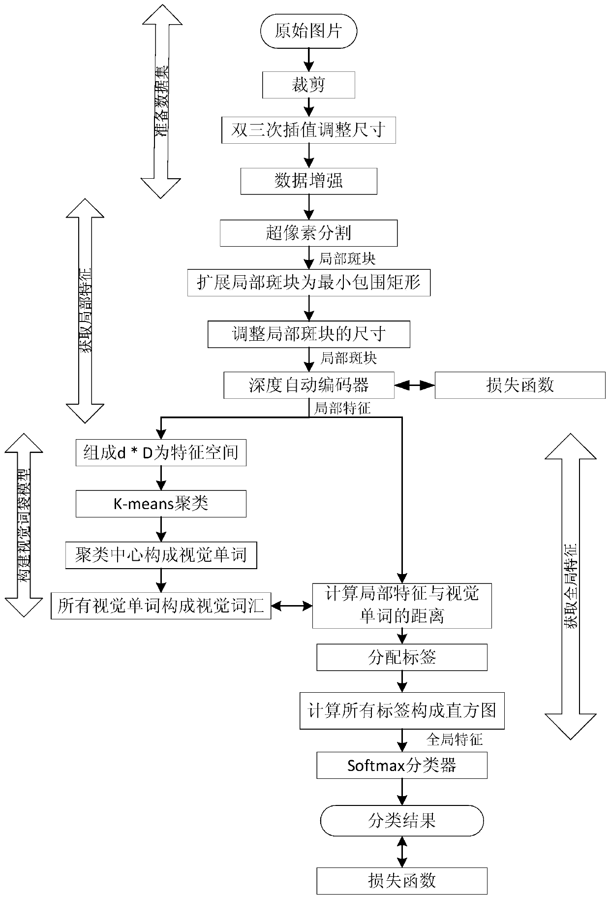
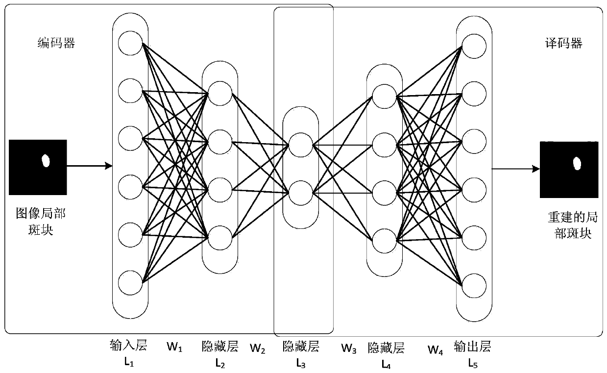
[0059] A lung nodule image classification method based on autoencoder to construct feature representation, such as figure 1 shown, including the following steps:

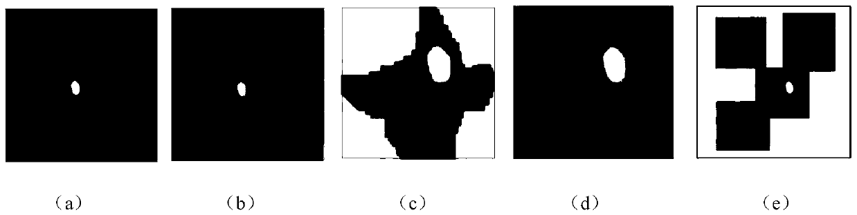
[0060] Step 1. Collect CT image data of pulmonary nodules According to the appearance of pulmonary nodules and their relationship with surrounding tissues and expert guidance, pulmonary nodules can be divided into good border type, pleural adhesion type, pleural...


PUM


Abstract

The invention provides a pulmonary nodule image classification method for constructing feature representation based on an automatic encoder, and relates to the technical field of computer vision. Themethod comprises the following steps: firstly, segmenting a pulmonary nodule image into local patches through superpixels; Transforming the patches into local feature vectors with a fixed length by using an unsupervised depth auto-encoder; Constructing visual vocabularies on the basis of the local features, and describing global features of the pulmonary nodule image through a visual word bag; Classifying pulmonary nodule types by using a softmax algorithm to complete the design of a model framework for representing pulmonary nodule image characteristics; Performing training by using the designed model framework and the ELCAP data set to obtain an automatic classification model for the pulmonary nodule images; And finally, carrying out pulmonary nodule image classification by using the obtained pulmonary nodule image classification model. According to the pulmonary nodule image classification method for constructing feature representation based on the automatic encoder provided by theinvention, the feature extraction capability of the pulmonary nodule classification model is improved, and the accuracy of pulmonary nodule automatic classification is improved.

Description

technical field [0001] The invention relates to the technical field of computer vision, in particular to a method for classifying pulmonary nodule images based on feature representations constructed by an automatic encoder. Background technique [0002] Lung cancer is one of the deadliest diseases in the world, accounting for approximately 20% of all cancers in 2016. Despite recent advances in diagnosis and treatment, the 5-year cure rate is only 18.2%. Notably, if patients are diagnosed early and properly treated, their chances of survival are greatly increased. Low-dose CT scans can reduce lung cancer deaths by 20 percent, according to the National Lung Screening report. The traditional diagnosis of pulmonary nodules mainly relies on the observation of doctors and experts, so there are some disadvantages: the diagnostic results are subjective; the workload is heavy and time-consuming; human vision is limited; doctors in remote areas cannot be popularized. With the devel...

Claims


Application Information

Patent Timeline
IPC IPC(8): G06K9/62G06K9/46G06T7/11
Inventor 毛克明王新琦李佳明李翰鹏常辉东尹贺
Owner NORTHEASTERN UNIV
Why Eureka
Try Eureka