
Gynecological tumor removing device

A technology for taking out devices and gynecological tumors, which is applied in the field of medical devices, can solve the problems of high probability of wound infection, slow recovery, and increased patient pain, and achieve the effects of convenient operation, reduced length, and prevention of secondary injuries

Inactive Publication Date: 2019-06-21
内蒙古民族大学附属医院
View PDF11 Cites 6 Cited by
  • Summary
  • Abstract
  • Description
  • Claims
  • Application Information

AI Technical Summary

Problems solved by technology

[0002] At present, when the gynecology department of the hospital performs uterine cavity tumor resection, there is no special machine for the removal of small tumors such as condyloma acuminata and papilloma. Usually, these small tumors are removed with forceps, and then small surgical scissors or surgery. Resection, the operation is very inconvenient. After the resection, most of the hands are stretched into the uterine cavity to grasp. This kind of operation method has a large incision in the uterine cavity, the patient recovers slowly after the operation, and the wound infection rate is high, which increases the pain of the patient and reduces the The work efficiency and work quality of medical staff need to be improved urgently

Method used


Image

  • Gynecological tumor removing device
  • Gynecological tumor removing device
  • Gynecological tumor removing device

Examples


Embodiment Construction

[0026] The present invention will be further described below in conjunction with the accompanying drawings.

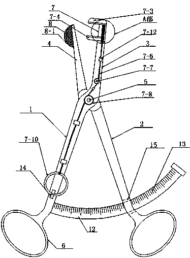
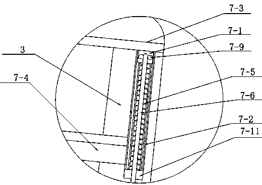
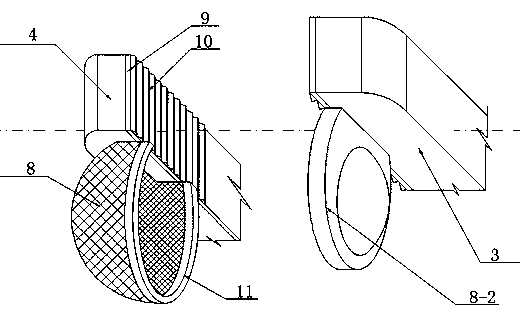
[0027] see as Figure 1-Figure 7 As shown, the technical solution adopted in this specific embodiment is: it comprises No. 1 pincer leg 1, No. 2 pincer leg 2, No. 1 pincer mouth 3, No. 2 pincer mouth 4, pin shaft 5, finger ring 6; No. 1 pincer leg 1 is arranged on the left side of the No. 2 tong leg 2, the upper end of the No. 1 tong leg 1 and the upper end of the No. 2 tong leg 2 are intersected, and the intersection is connected by a pin shaft 5. The No. 1 tong leg 1 and the No. 2 tong leg The connection mode and working principle of the legs 2 and the pin shaft 5 are the same as those of the two arms of the medical tweezers in the prior art; The upper end of the No. 2 pincer leg 2 is fixedly provided with a No. 2 pincer mouth 4; the No. 1 pincer leg 1 and the No. 1 pincer mouth 3, as well as the No. 2 pincer leg 2 and the No. 2 pincer mouth 4 are all made of medica...


PUM


Abstract

The invention discloses a gynecological tumor removing device, and relates to the technical field of medical instruments. A first pincer leg is arranged on the left side of a second pincer leg, the upper end of the first pincer leg and the upper end of the second pincer leg are intersected, intersected portions are rotatably connected by a pin shaft, a first pincer nozzle is fixedly arranged at the upper end of the first pincer leg, a second pincer nozzle is fixedly arranged at the upper end of the second pincer leg, the first pincer leg and the first pincer nozzle are integrated, the second pincer leg and the second pincer nozzle are integrated, finger rings are arranged at the lower ends of the first pincer leg and the second pincer leg, the gynecological oncology removing device comprises a tumor cutting device and a tumor removing device, the tumor cutting device is fixedly arranged on the front side wall of the first pincer nozzle and can help medical staff to cut a tumor in a uterine cavity of a patient, surgical operation is more conveniently implemented, the medical staff does not need to manually enter the uterine cavity of the patient and grab the tumor in the uterine cavity, the length of an incision of the uterine cavity is reduced, and secondary harm to the patient is prevented.

Description

technical field [0001] The invention relates to the technical field of medical devices, in particular to a device for removing gynecological tumors. Background technique [0002] At present, when the gynecology department of the hospital performs uterine cavity tumor resection, there is no special machine for the removal of small tumors such as condyloma acuminata and papilloma. Usually, these small tumors are removed with forceps, and then small surgical scissors or surgery. Resection, the operation is very inconvenient. After the resection, most of the hands are stretched into the uterine cavity to grasp. This kind of operation method has a large incision in the uterine cavity, the patient recovers slowly after the operation, and the wound infection rate is high, which increases the pain of the patient and reduces the The work efficiency and work quality of medical staff need to be improved urgently. Contents of the invention [0003] The object of the present invention...

Claims


Application Information

Patent Timeline
Patent Type & Authority Applications(China)
IPC IPC(8): A61B17/3205A61B17/285A61B17/42A61B10/04
Inventor 包克勇张雪陶晓玉许永慧于淑红禹彬张莉肖雨竹李娟毕秀娟
Owner 内蒙古民族大学附属医院
Learn More