A nasopharyngeal carcinoma focus segmentation model training method and segmentation method based on deep learning

A segmentation model and deep learning technology, which is applied in the field of medical image processing, can solve problems such as poor results, and achieve the effect of improving the segmentation effect and accurate automatic segmentation

Inactive Publication Date: 2019-06-21
SOUTH CHINA UNIV OF TECH
View PDF4 Cites 16 Cited by
  • Summary
  • Abstract
  • Description
  • Claims
  • Application Information

AI Technical Summary

Problems solved by technology

[0006] The purpose of the present invention is to overcome the shortcomings and deficiencies in the prior art, to provide a deep learning-based nasopharyngeal carcinoma lesion segmentation model training method and a segmentation method, the model obtained by the segmentation model training method is a...

Method used


Image

  • A nasopharyngeal carcinoma focus segmentation model training method and segmentation method based on deep learning
  • A nasopharyngeal carcinoma focus segmentation model training method and segmentation method based on deep learning
  • A nasopharyngeal carcinoma focus segmentation model training method and segmentation method based on deep learning

Examples


Embodiment

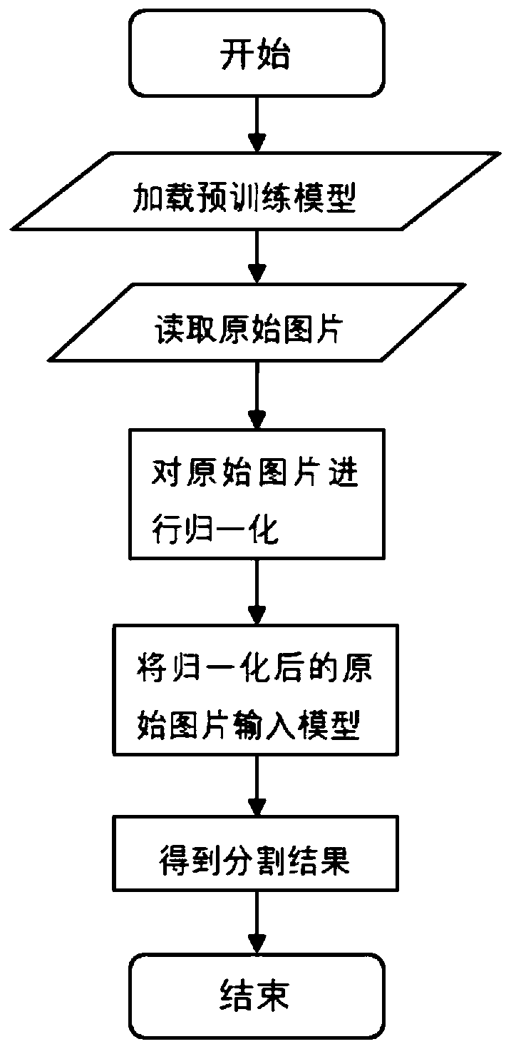
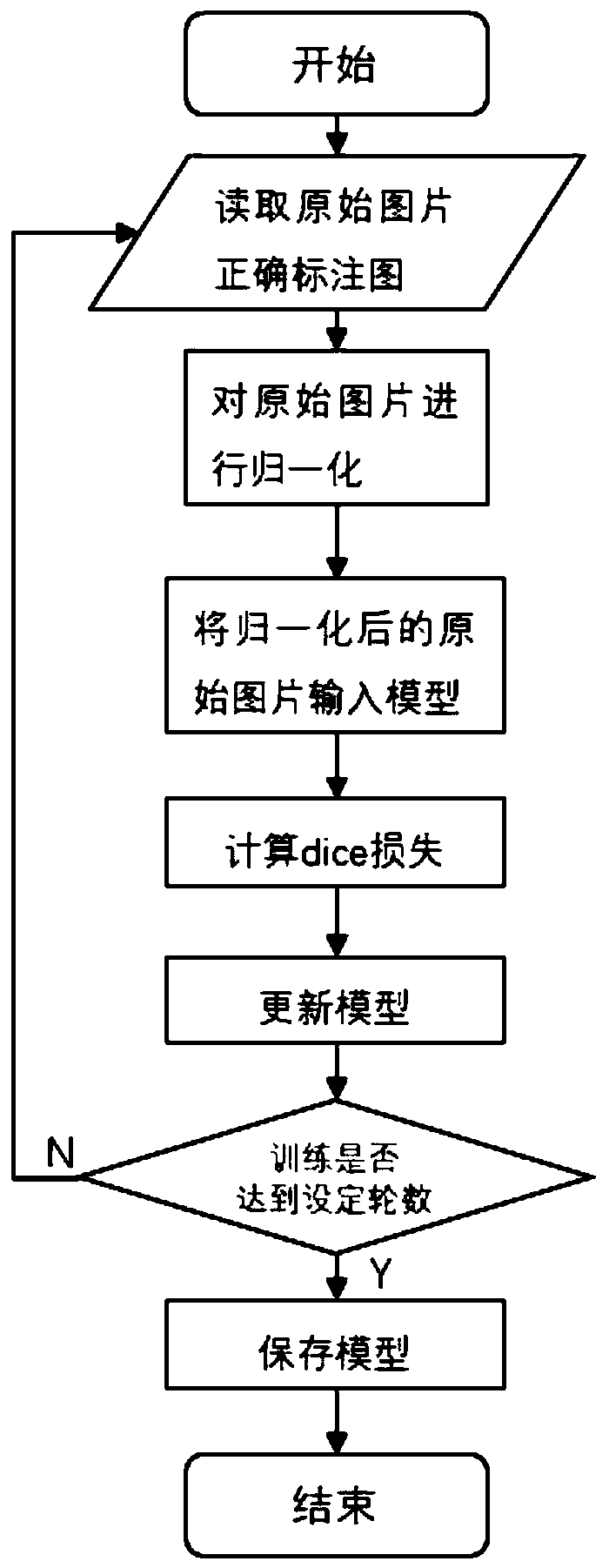
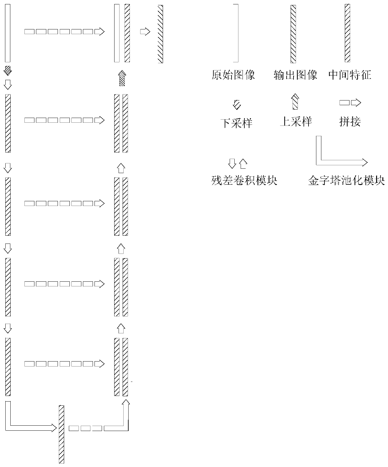
[0061] Such as Figure 1 to Figure 5 As shown, a kind of deep learning-based nasopharyngeal carcinoma lesion segmentation method of the present invention comprises the following steps:

[0062] I. Load the trained segmentation model into the convolutional neural network model;

[0063] II. read the nasopharyngeal carcinoma MRI original image, the nasopharyngeal carcinoma MRI original image is a single-channel grayscale image;

[0064] III. Normalizing the original MRI image of nasopharyngeal carcinoma to obtain the MRI image of nasopharyngeal carcinoma;

[0065] IV. Input the MRI image of nasopharyngeal carcinoma into the convolutional neural network model to obtain the lesion probability map of the MRI image of nasopharyngeal carcinoma;

[0066] V. Binarize the lesion probability map of the nasopharyngeal carcinoma MRI image to obtain a lesion segmentation map.

[0067] Among them, binarizing the lesion probability map of the nasopharyngeal carcinoma MRI image to obtain th...


PUM


Abstract

The invention provides a nasopharyngeal carcinoma focus segmentation model training method and segmentation method based on deep learning, and the method comprises the following steps: I, loading a trained segmentation model into a convolutional neural network model; II, reading the nasopharyngeal carcinoma MRI original image; III, normalizing the nasopharyngeal carcinoma MRI original image to obtain a nasopharyngeal carcinoma MRI image; IV, inputting the nasopharyngeal carcinoma MRI image into the convolutional neural network model to obtain a focus probability map of the nasopharyngeal carcinoma MRI image; And V, binarizing the focus probability map of the nasopharyngeal carcinoma MRI image to obtain a focus segmentation map. The invention further provides a nasopharyngeal carcinoma focus segmentation model training method. The model obtained through the nasopharyngeal carcinoma focus segmentation model training method based on deep learning is applied to the focus segmentation method, and the problem of poor effect caused by small image segmentation in the nasopharyngeal carcinoma MRI image can be effectively solved, so that the segmentation effect of the target small image is improved, and the nasopharyngeal carcinoma focus is segmented more accurately and automatically.

Description

technical field [0001] The present invention relates to the technical field of medical image processing, and more specifically, to an automatic segmentation model training method and segmentation method for nasopharyngeal carcinoma lesions based on deep learning. Background technique [0002] Magnetic Resonance Imaging (MRI) uses static magnetic field and radio frequency magnetic field to image human tissue. During the imaging process, it can obtain clear images with high contrast without ionizing radiation or contrast agent. It can reflect the abnormalities and early lesions of human organs from the inside of human molecules. Magnetic resonance imaging can provide information on the physiological state of human organs, and it has the ability to characterize different tissues of the same density and different chemical structures of the same tissue through image display. [0003] Nasopharyngeal carcinoma (NPC) refers to malignant tumors that occur on the top and side walls o...

Claims


Application Information

Patent Timeline
IPC IPC(8): G06T7/11G06T7/136G06N3/04G06N3/08
Inventor 夏康力田翔晋建秀
Owner SOUTH CHINA UNIV OF TECH
Why Eureka
Try Eureka