A minimally invasive fusion device

A cage and mesh bag technology, applied in the direction of spinal implants, etc., can solve the problems that the cage does not have mechanical support, restricts the space for bone grafting, and affects the fusion effect.

Active Publication Date: 2021-07-30
NINGBO HICREN BIOTECH
View PDF9 Cites 0 Cited by
  • Summary
  • Abstract
  • Description
  • Claims
  • Application Information

AI Technical Summary

Problems solved by technology

Although the height of this mechanism can be raised, it is not scalable in the width direction. If you want to pass through the slender working channel of the endoscope, the width must be narrow, and the length is limited by the size of the vertebral body. The final contact area with the upper and lower endplates Relatively small, resulting in excessive local pressure of the vertebral body, which will cause the fusion cage to sink into the interior of the vertebral body for a long time
[0008] It can be seen from the above that in the existing intervertebral fusion surgery, some fusion cages do not have good mechanical support and need to be filled with self-coagulating materials to achieve the purpose of mechanical support. The amorphous shape of self-coagulating materials often leads to poor surgical results , and then a second operation is required, which brings a heavy burden to the patient; in the mechanical structure of the minimally invasive fusion device in the existing patent, it can only realize the variable height direction or the variable width direction. The two are incompatible in terms of vertebral body contact area, and the mechanical structure limits the space for bone grafting, which affects the fusion effect; in addition, most of the fusion cages used in intervertebral fusion surgery are fixed in shape, large in size, and difficult to implant. Larger access channels are required, and the patient is more injured

Method used


Image

  • A minimally invasive fusion device
  • A minimally invasive fusion device
  • A minimally invasive fusion device

Examples


Embodiment 1

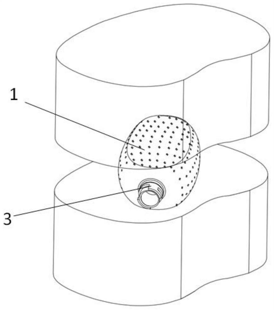
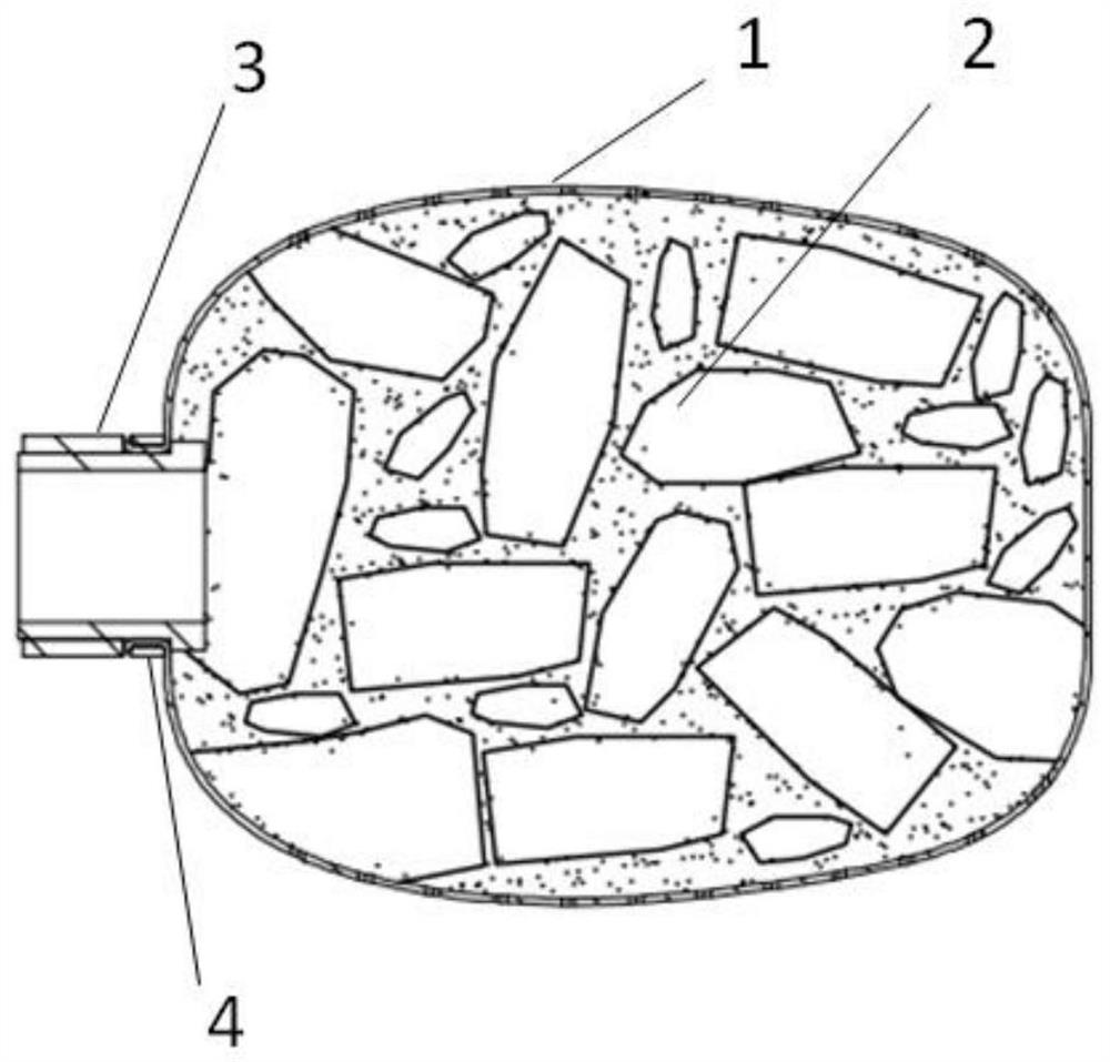
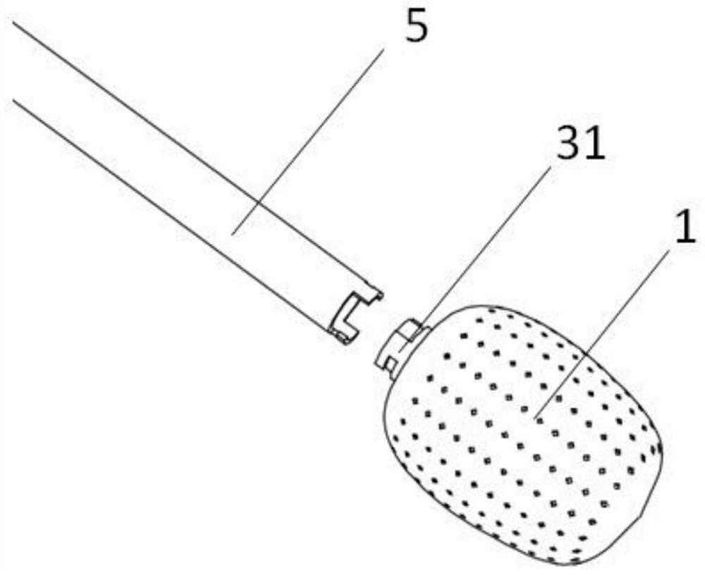
[0076] Such as figure 2 As shown, a minimally invasive fusion device includes a mesh bag 1 and a filling material 2. A connector 3 is provided at the proximal end of the mesh bag 1, and the connector 3 is extruded and connected to the mesh bag 1 through a collar 4. Such as image 3 As shown, the mesh bag 1 is connected to the connecting pipe 5 through the connecting head 3, and the mesh bag 1 is sent into the intervertebral space along the working channel through the connecting pipe 5, as shown in FIG. figure 1 shown; after reaching the working position, such as Figure 7 and 8 As shown, fill the filling tube 6 with the filling material 2 times, and use the inner push rod 7 to fill it into the inside of the mesh bag 1 until the mesh bag 1 expands to fit the contact surface of the upper and lower vertebral bodies. After filling, such as figure 2 As shown, the filling material 2 can seal the inner hole of the connector 3 and play a role of self-sealing. The inner push rod...

Embodiment 2

[0082] Such as figure 2 As shown, a minimally invasive fusion device includes a mesh bag 1 and a filling material 2. A connecting head 3 is provided at the proximal end of the mesh bag 1, and the connecting head 3 is extruded and connected to the mesh bag 1 through a collar 4. Bone filling materials 2 with different structures and particle sizes are preloaded in the filling tube 6 according to the ratio. Such as image 3 and 4 As shown, the mesh bag 1 is connected to the connecting tube 5 through the connecting head 3, and the mesh bag 1 is sent into the intervertebral space along the working channel through the connecting tube 5. After arriving at the work location, such as Figure 5 and 6 As shown, the filling pipe 6 enters the mesh bag 1 along the connecting pipe 5, and the filling material 2 in the filling pipe 6 is pushed to the inside of the mesh bag 1 by using the inner push rod 7 until the mesh bag 1 is fully expanded to the point where the upper and lower vertebr...

Embodiment 3

[0089] (The fuser has a liner without a sealing device) such as Figure 11 As shown, a minimally invasive fusion device includes a foldable mesh bag 1, a filling material 2 and a liner core 9, an outlet 10 is provided in the middle, and a connector 3 is provided at the proximal end, the mesh bag 1 and the liner The proximal end of the core 9 is extruded and connected through a collar 4 , the connecting head 3 is extruded connected with the lining core 9 , and the distal end is extruded connected with the lining core 9 through another collar 4 . The connecting tube 5 can be connected with the connector 3 of the fusion device, and the fusion device is sent into the intervertebral space along the working channel. After reaching the working position, as Figure 7 and 8 As shown, the filling material 2 is filled in the filling tube 6 several times. Use the inner push rod 7 to inject the filling material 2 into the net bag 1 through the outlet 10, and the net bag 1 can expand to f...


PUM


Abstract

The invention discloses a minimally invasive fusion device, which comprises a foldable or telescopic mesh bag, and a connecting head which can communicate with the outside is arranged at the proximal end of the mesh bag; material; and / or a tubular lining core is arranged inside the mesh bag, an outlet is opened on the lining core, and its proximal end communicates with the connector, and the far end is fixed to the far end of the mesh bag; and / or the internal distal position of the lining core A sealing device is provided; the inner hole or outlet of the connector is blocked by changing the position of the filling material injected into the mesh bag, and / or the sealing device resets to block the outlet under the action of its own elasticity. The minimally invasive fusion device provided by the present invention can be self-sealed after the net bag is filled, and the sealing treatment of the net bag can prevent the bone filling material from leaking out and affect the fusion effect, and the automatic sealing simplifies the operation, shortens the operation time, and improves the operation efficiency. safety.

Description

technical field [0001] The invention relates to the field of spinal intervertebral fusion, in particular to a minimally invasive fusion device. Background technique [0002] Degenerative diseases of the spine and its structural damage are an important cause of neck, shoulder, waist, and leg pain, sensory and motor function impairment or even loss. In the 1950s, Cloward first proposed Posterior Lumbar Fusion (PLIF), which has developed into one of the basic procedures of spinal surgery today. In 1986, Badgy and Kuslich designed an intervertebral fusion device (Cage) suitable for the human body, namely the BAK system. Since then, intervertebral bone graft fusion technology has been greatly developed and has become a basic surgical method for the treatment of spinal degenerative diseases and structural injuries. [0003] The principle of the intervertebral fusion device is centered on the diseased intervertebral space. After the intervertebral fusion device is implanted, the ...

Claims


Application Information

Patent Timeline
Patent Type & Authority Patents(China)
IPC IPC(8): A61F2/44
CPCA61F2/4455
Inventor 陈琼威卓清山吕世文杨洋张鹏云
Owner NINGBO HICREN BIOTECH
Why Eureka
Try Eureka