Tissue lockable connecting structures

a technology of tissue locking and connecting structures, which is applied in the direction of prosthesis, dental surgery, catheters, etc., can solve the problems of soft tissue infection and other complications, weak and insufficient bio-sealing connection between the skin and the device, and disrupt the protective barrier, etc., to achieve strong bio-connection, inhibit tissue and skin infection, and improve mechanical connections

Inactive Publication Date: 2006-08-01
EAST CAROLINA UNIVERISTY
View PDF25 Cites 138 Cited by
  • Summary
  • Abstract
  • Description
  • Claims
  • Application Information

AI Technical Summary

Benefits of technology

[0007]In certain embodiments, the present invention can provide locked connecting structures (LCS) which engage with the localized tissue and provide improved mechanical connections at a junction layer located between the outer surface of the device and the tissue locking structures mounted thereto. The mechanical connection protects the bio-sealing connection and the intact bio-sealing connection, in turn, inhibits tissue and skin infection, even in long-term indwelling applications. The locked connecting structures can be used for a number of medical applications including on percutaneous access devices. The locked connecting structures are adapted to provide a sufficiently strong bio-connection and with sufficiently strong structural support so as to inhibit the disruption of the bio-sealing connection and enhance the survivability of the bio-sealing connection during normal life activities even during long-term indwelling applications.

Problems solved by technology

Many implantable artificial organs and foreign devices require skin penetration which can disrupt the protective barrier typically provided by the skin under natural conditions.
One common problem associated with PADs, implanted on a long-term basis, is that the bio-sealing connection between the skin and the device can be weak and not sufficiently strong to be able to resist detaching or fracturing during the stress introduced onto the skin during normal life activities.
Unfortunately, a break in the bio-seal connection can lead to soft tissue infection and other complications.
However, and unfortunately, the configuration or location of the flange typically limited its ability to provide suitable connective assistance.
Unfortunately, each of these types of devices may cause infection, skin downgrowth, and other complications.
Further, the bio-sealing connections proposed by these conventional PADs may be too weak to maintain the integrity of the bio-sealing connection during normal life activities, particularly when implanted for chronic or long-term indwelling periods of time.

Method used


Image

  • Tissue lockable connecting structures
  • Tissue lockable connecting structures
  • Tissue lockable connecting structures

Examples


example 1

Structure, Material, and Method

[0143]This study was approved by the Animal Care and Use Committee (East Carolina University School of Medicine), and the care and handling of animals were in accord with national Institutes of Health guidelines.

[0144]The LPD-I device tested (“locking percutaneous device”) had two portions. The solid clear plexiglass dome was a prototype simulating any medically necessary device. The dome diameter studied ranged from 2.3–2.7 cm, the thickness at the edge was approximately 0.7 cm, and at its center 1.1 cm. The second portion of the LPD-I was the connecting structure that included two parts: the skin connector and the subcutaneous connector. FIGS. 11A and 11B are digital images of the studied device.

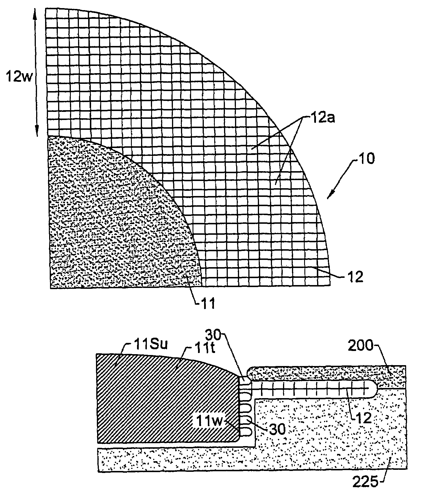
[0145]The skin connector was a mesh collar made from polyester net with 72 eyes / cm2. The diameter of the warp and weft was approximately 0.2 mm. The mesh was structured so that the warp and the weft did not slide over each other, thereby fixing the pore size ...

example 2

Structure, Material, and Method

[0157]Two types of LPD-II devices were tested. Type 1 had two components, a dome and a skin connector (but without the underlying skin stop collar). The Type 2 device had an extra structure, the SSC (see FIG. 10). The dome was constructed from solid clear plexiglass with a thickness of 0.4 cm and a diameter of 2.3–3.5 cm. The skin connector or mesh collar surrounded the dome. It was made from polyester net with 72 eyes / cm2 and about 0.2 mm diameter of the warp and weft. Its width was approximately 1.3 cm. The SSC was positioned just beneath the mesh collar and was made from stainless steel mesh with 64 eyes / cm2. The wire had a diameter of 0.5 mm and width of approximately 0.4 cm.

[0158]The mesh collar of the LPD-II without the SSC (type 1) was attached to the bottom edge of the dome by heating. The mesh collar and SSC (LPD-II type 2) were fixed to the lower edge of the dome simultaneously first by heating and then by application of a layer of thick plex...


PUM


Abstract

Percutaneous skin access devices include a plurality of locked connecting units mounted to the exterior surface of an implantable medical object which, in position, is configured to penetrate the skin of a subject. The locked connecting units may be mounted directly onto the desired surface of the exterior of the device or may be held on a substrate sheet, which is mounted to the exterior surface of the device. In position, the locked connecting units engage with soft tissue which can include the skin to form a bio-junction layer which includes mechanical and bio-sealing connection between the device body and the soft tissue. The configuration at the bio-junction layer secures the medical object in location in the subject even for long-term indwelling applications in a manner, which inhibits soft tissue infection.
The locked connecting units may be rigid or semi-rigid for longer-term indwelling applications, and semi-rigid and/or resilient for shorter term indwelling applications. The locked connecting units may take on the form of rings, hooks, or loops having aperture or gap width/length sizes of from about 0.2–4 mm. The rings, loops, or hooks may connect with any soft tissue including skin as well subcutaneous tissue. The rings, hooks, or loops may be released from the skin/tissue without requiring surgical cutting procedures.
The locked connecting units may be configured as a semi-rigid mesh collar arranged about the primary body providing access to the subject such that it resides in the subject and engages with the skin (epidermal/dermal layer). The mesh collar can be described as a particular type of ring or loop structure as the mesh defines the gap provided in individual loop configurations. The mesh collar may be used alone, or in combination with the loops, rings, or hooks. A skin stop collar having increased rigidity may be disposed under the mesh collar.

Description

RELATED APPLICATIONS[0001]This application claims the benefit of priority from PCT Application No. US0 / 32606 filed Nov. 30, 2000 which claimed benefit of priority from U.S Provisional Application Ser. No. 60 / 244,735 filed Oct. 31, 2000, the contents of which are hereby incorporated by reference as if recited in full herein.FIELD OF THE INVENTION[0002]The invention relates to tissue locking structures and implantable percutaneous barrier devices for providing a way to enter through the skin of a subject safely and chronically for artificial or foreign objects such as wires, tubing, bars, plates, containers, and also relates to bioconnection structures useful for other devices such as skeletal pins, dental implants, or prostethic devices.BACKGROUND OF THE INVENTION[0003]Generally stated, skin functions to shield internal tissues from the environment and to protect against infection. Many implantable artificial organs and foreign devices require skin penetration which can disrupt the p...

Claims


Application Information

Patent Timeline
Patent Type & Authority Patents(United States)
IPC IPC(8): A61F2/10A61C8/00A61F2/00A61F2/30A61F2/50A61F2/60A61F2/68A61F2/70A61F2/72A61F2/76A61F2/78A61M25/02A61M25/04
CPCA61M25/04A61F2/60A61F2/68A61F2/78A61C8/0009A61F2/72A61F2002/0068A61F2002/0086A61F2002/30467A61F2002/30914A61F2002/3093A61F2002/704A61F2002/705A61F2002/7615A61F2002/7881A61F2002/7887A61F2220/0083A61F2310/00011A61M25/02A61F2/30749
Inventor YU, CHANGHARRIS, GLENN
Owner EAST CAROLINA UNIVERISTY
Why Eureka
Try Eureka