Fundus blood vessel image classification method and device, equipment and storage medium

A blood vessel image and classification method technology, applied in the field of fundus blood vessel image classification method, device, equipment and storage medium, to achieve the effect of ensuring robustness and classification accuracy

Active Publication Date: 2019-10-18
腾讯医疗健康(深圳)有限公司
View PDF7 Cites 14 Cited by
  • Summary
  • Abstract
  • Description
  • Claims
  • Application Information

AI Technical Summary

Problems solved by technology

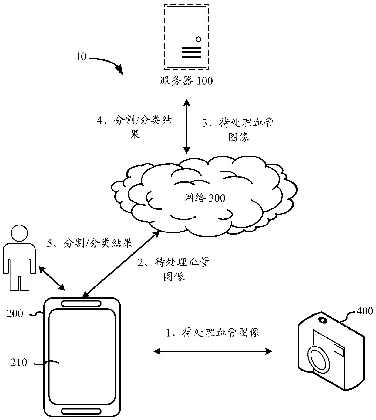
[0004] There is no effective solution compatible with related technologies to realize the automation of multiple tasks of vessel segmentation and classification

Method used


Image

  • Fundus blood vessel image classification method and device, equipment and storage medium
  • Fundus blood vessel image classification method and device, equipment and storage medium
  • Fundus blood vessel image classification method and device, equipment and storage medium

Examples


Embodiment Construction

[0062] In order to make the purpose, technical solution and advantages of the present invention clearer, the present invention will be described in further detail below in conjunction with the accompanying drawings, and the described embodiments should not be considered as limiting the present invention, and those of ordinary skill in the art do not make any All other embodiments obtained under the premise of creative labor belong to the protection scope of the present invention.

[0063] In the following description, references to "some embodiments" describe a subset of all possible embodiments, but it is understood that "some embodiments" may be the same subset or a different subset of all possible embodiments, and Can be combined with each other without conflict.

[0064] Unless otherwise defined, all technical and scientific terms used herein have the same meaning as commonly understood by one of ordinary skill in the technical field of the invention. The terms used herei...


PUM


Abstract

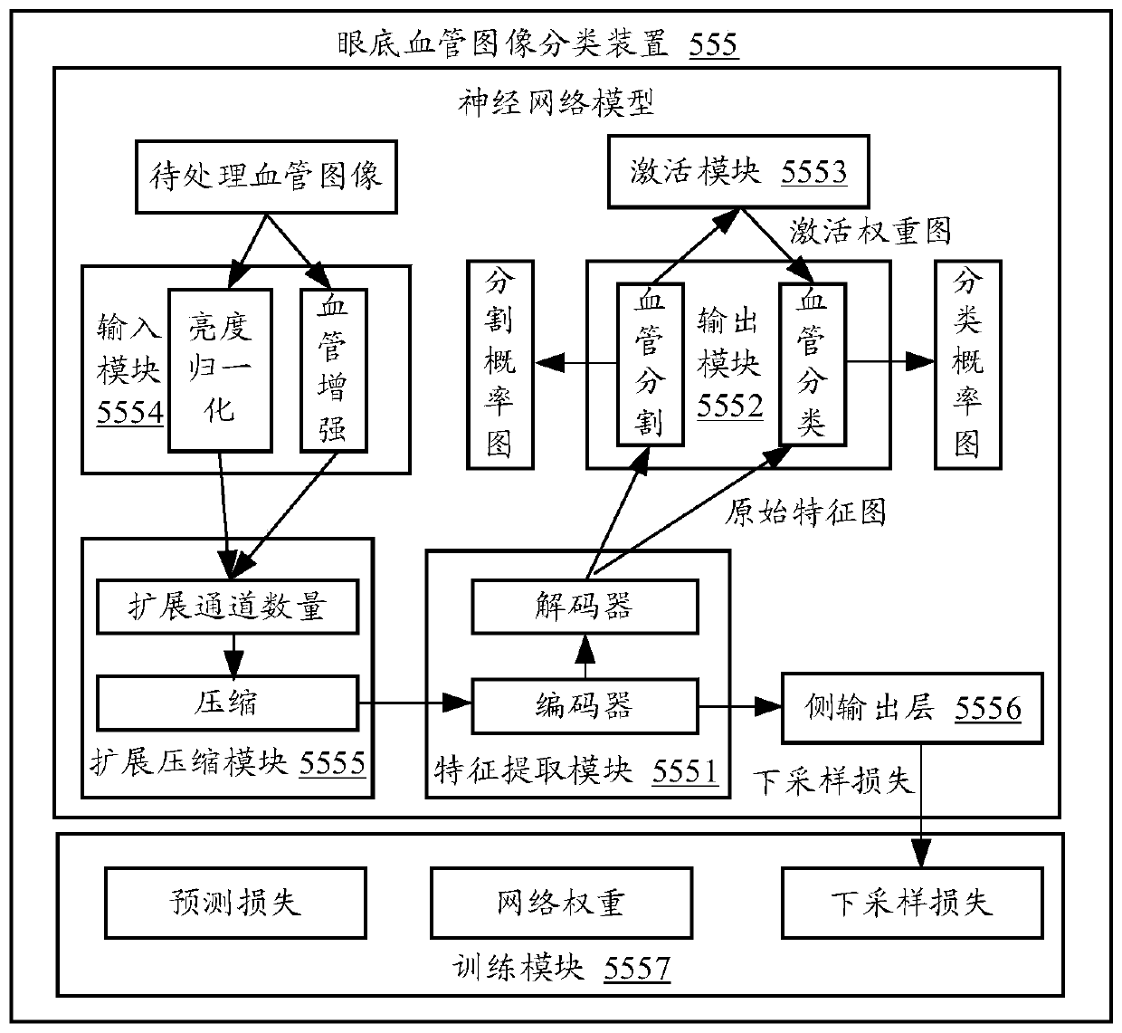
The invention provides a fundus blood vessel image classification method and device, equipment and a storage medium. The method comprises the following steps: extracting an original feature map from afundus blood vessel image; based on the features of each pixel point in the original feature map, determining a probability value of each pixel point belonging to a blood vessel in the fundus blood vessel image to form a blood vessel segmentation probability map of the fundus blood vessel image; distributing a corresponding weight to each pixel point based on the distribution condition of the probability values corresponding to each pixel point in the blood vessel segmentation probability graph; fusing the weight of each pixel point in the fundus blood vessel image with the feature of the corresponding pixel point in the original feature map to obtain a fused feature map; and based on the features of each pixel point in the fusion feature map, determining a probability value that each pixel point in the fundus blood vessel image belongs to an artery blood vessel and a vein blood vessel, and forming a blood vessel classification probability map of the fundus blood vessel image. According to the invention, blood vessel segmentation and blood vessel classification can be automatically realized with high precision.

Description

technical field [0001] The invention relates to medical image processing technology, in particular to a fundus blood vessel image classification method, device, equipment and storage medium. Background technique [0002] Segmentation (i.e. identifying blood vessels from the background) and classification (i.e. classifying blood vessels) in images is of great significance in clinical diagnosis and treatment. [0003] Taking fundus blood vessels as an example, the fundus is the only area in the human body where blood vessels can be directly observed without intervention. Many systemic diseases and cardiovascular and cerebrovascular diseases will affect the shape of fundus blood vessels, and have different effects on the formation of arteries and veins. For example, clinical studies have confirmed that the reduction of the fundus arteriovenous width ratio will increase the risk of stroke; the narrowing of the fundus arteries is related to the development of hypertension and dia...

Claims


Application Information

Patent Timeline
Patent Type & Authority Applications(China)
IPC IPC(8): G06K9/62G06K9/46G06T7/10
CPCG06T7/10G06T2207/10024G06T2207/30101G06T2207/30041G06T2207/20084G06T2207/20076G06T2207/20081G06V10/40G06F18/2415G06F18/253
Inventor 余双
Owner 腾讯医疗健康(深圳)有限公司
Why Eureka
Try Eureka