
Automatic division method for pulmonary parenchyma of CT image

A technology for automatic segmentation of CT images, applied in image analysis, image enhancement, image data processing, etc., can solve problems such as ineffective segmentation of lung parenchyma in CT images

Inactive Publication Date: 2015-10-21
HEBEI UNIVERSITY
View PDF2 Cites 36 Cited by
  • Summary
  • Abstract
  • Description
  • Claims
  • Application Information

AI Technical Summary

Problems solved by technology

[0005] The purpose of the present invention is to provide a method for automatic segmentation of lung parenchyma in CT images to solve the problem that the existing lung parenchyma segmentation methods cannot effectively segment the lung parenchyma of CT images in complex situations

Method used


Image

  • Automatic division method for pulmonary parenchyma of CT image
  • Automatic division method for pulmonary parenchyma of CT image
  • Automatic division method for pulmonary parenchyma of CT image

Examples


Embodiment Construction

[0046] The software and hardware condition of computer used in the embodiment of the present invention is: Dual-Core CPU E5800 3.20GHz, graphics card is NVIDIA GeForce GT 430, memory 2.0GB, operating system is Window 2007, software programming language uses MATLAB. This invention is funded by the National Natural Science Foundation of China (No. 61375075).

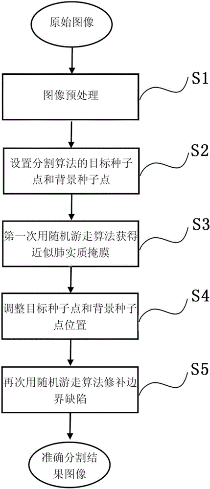
[0047] Such as figure 1 Shown, the present invention comprises the steps:

[0048] Step S1, image preprocessing.

[0049] This step preprocesses the original CT image, mainly including Gaussian smoothing and denoising, and uses Otsu threshold segmentation technology to binarize the image to obtain the target mask and background mask. details as follows:

[0050] (1) Gaussian smoothing denoising: use 3×3 Gaussian template to filter the image to achieve the purpose of reducing noise.

[0051] (2) Obtain the target mask and background mask: use the Otsu threshold segmentation technique (maximum inter-class variance meth...


PUM


Abstract

The invention provides an automatic division method for pulmonary parenchyma of a CT image. According to the automatic division method, the CT is divided through carrying out a random migration algorithm for two times to obtain the accurate pulmonary parenchyma; in the first time, the random migration algorithm is used for dividing to obtain a similar pulmonary parenchyma mask; and in the second time, the random migration algorithm is used for repairing defects of the periphery of a lung and dividing to obtain an accurate pulmonary parenchyma result. Seed points, which are set by adopting the random migration algorithm to divide, are rapidly and automatically obtained through methods including an Otsu threshold value, mathematical morphology and the like; and manual calibration is not needed so that the working amount and operation time of doctors are greatly reduced. According to the automatic division method provided by the invention, a process of 'selecting the seed points for two times and dividing for two times' is provided and is an automatic dividing process from a coarse size to a fine size; and finally, the dependence on the selection of the initial seed points by a dividing result is reduced, so that the accuracy, integrity, instantaneity and robustness of the dividing result are ensured. The automatic division method provided by the invention is funded by Natural Science Foundation of China (NO: 61375075).

Description

technical field [0001] The invention relates to a method for segmenting lung parenchyma, in particular to an automatic segmenting method for lung parenchyma in CT images based on a random walk algorithm. Background technique [0002] At present, lung cancer is one of the malignant tumors with the fastest increasing morbidity and mortality and the greatest threat to human health and life. According to the latest research data released by the World Health Organization in May 2014, lung cancer ranks fifth among the top ten causes of death in the world and ranks first in cancer deaths. In my country, due to the large number of smokers and serious air pollution, the mortality rate of lung cancer has increased significantly, with an average annual rate of 4.4%. Since 1996, lung cancer has become the number one killer of cancer patients in my country. If lung cancer can be diagnosed and treated at an early stage, its five-year survival rate can reach 40% to 70%. [0003] Computed...

Claims


Application Information

Patent Timeline
Patent Type & Authority Applications(China)
IPC IPC(8): G06T7/00
CPCG06T2207/10081G06T2207/20061
Inventor 王兵顾潇蒙顾力栩张欣田学东
Owner HEBEI UNIVERSITY
Learn More