

Method and system for recognizing lattice degeneration and holes in wide area fundus images based on deep learning
A deep learning and fundus image technology, applied in the field of medical image processing, can solve problems such as undetectable and evaluation, time-consuming inspection, visual impairment, etc., and achieve the effects of reducing training expenditure, efficient disease assessment, and fast photography speed
- Summary
- Abstract
- Description
- Claims
- Application Information
AI Technical Summary
Problems solved by technology
Method used
Image

Examples
Embodiment 1
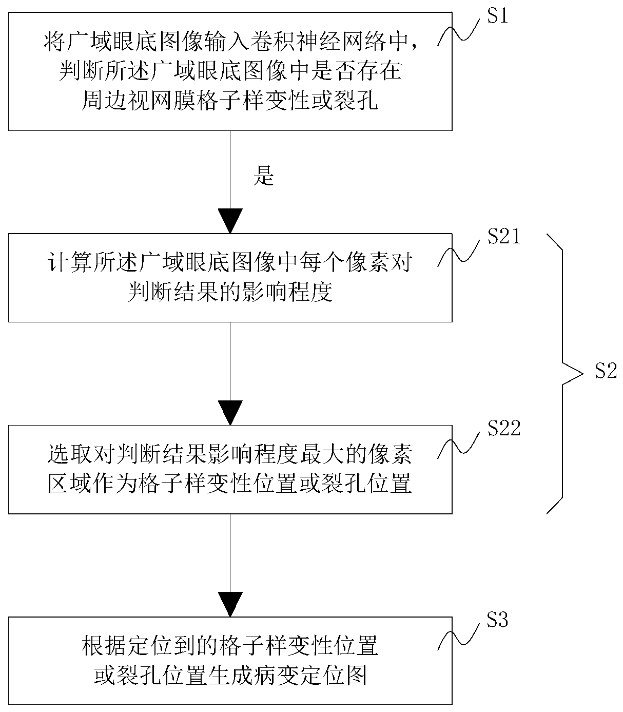
[0041] Such as figure 1 As shown, the present embodiment provides a method for identifying grid change holes in a wide-area fundus image based on deep learning, including the following steps:
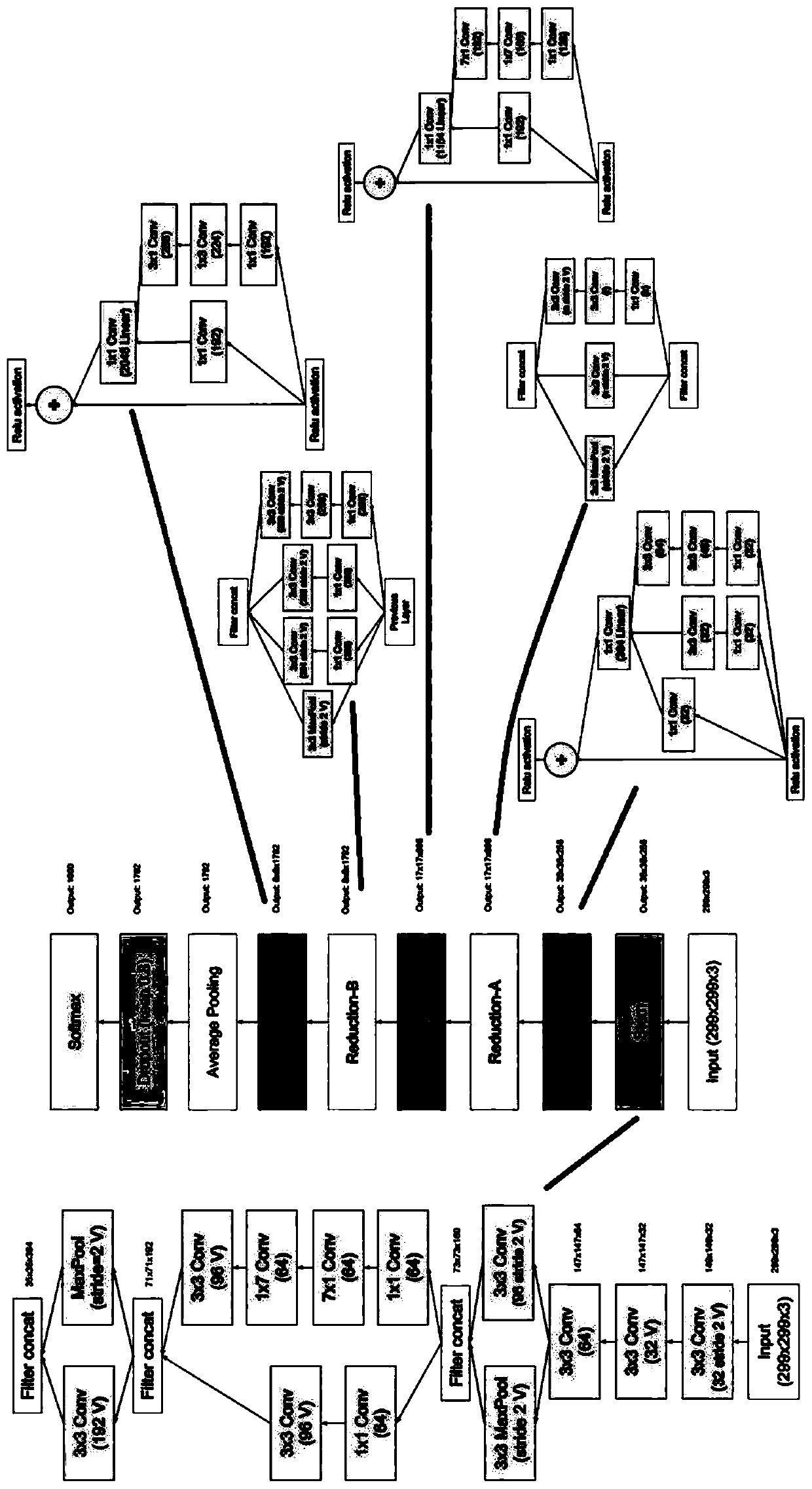
[0042] S1. Input the wide-area fundus image into the convolutional neural network, and judge whether there is lattice degeneration or hole in the peripheral retina in the wide-area fundus image;
[0043] S2. When it is judged that there is lattice degeneration or hole in the peripheral retina in the wide-area fundus image, the location of the lattice degeneration or the hole in the wide-area fundus image is located using a significant region algorithm.
[0044] In step S1, the convolutional neural network can be used to accurately and efficiently analyze the wide-area fundus image to determine whether there is a lesion on the image; in step S2, the salient area algorithm (Saliency Map) can be used to locate the lesion on the image. This can assist ophthalmologists to interpret patients...
Embodiment 2
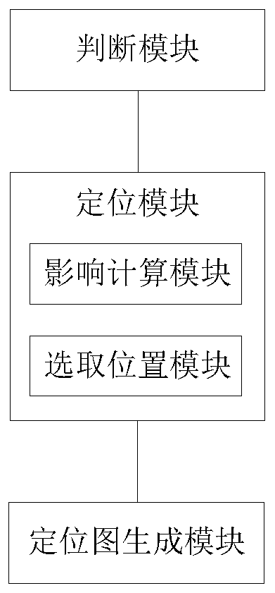
[0073] Such as image 3 As shown, this embodiment provides a system for identifying grid change holes in wide-area fundus images based on deep learning, including:
[0074] A judging module, configured to input the wide-area fundus image into the convolutional neural network, and judge whether there is a lattice-like degeneration or hole in the peripheral retina in the wide-area fundus image;
[0075] The positioning module is used to locate the grid-like degeneration or hole in the wide-area fundus image by using the salient area algorithm when it is judged that there is a grid-like degeneration or hole in the surrounding retina in the wide-area fundus image;
[0076] The judging module can accurately and efficiently analyze the wide-area fundus image through the convolutional neural network, and judge whether there is a lesion on the image; the positioning module can locate the location of the lesion on the image through the salient area algorithm (Saliency Map), which can a...
PUM
 Abstract
Description
Claims
Application Information